

Deoxycytidine kinase augments ATM-Mediated DNA repair and contributes to radiation resistance
- PMID: 25101980
- PMCID: PMC4125169
- DOI: 10.1371/journal.pone.0104125
Deoxycytidine kinase augments ATM-Mediated DNA repair and contributes to radiation resistance
Abstract
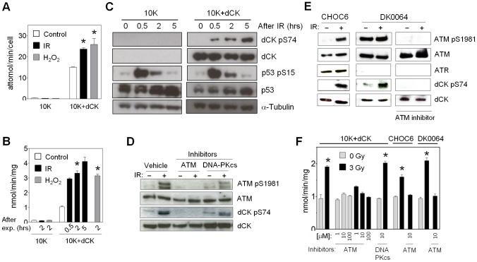
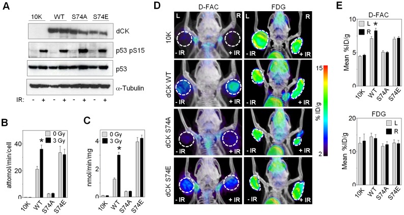
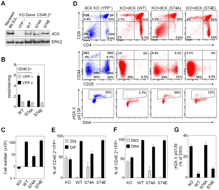
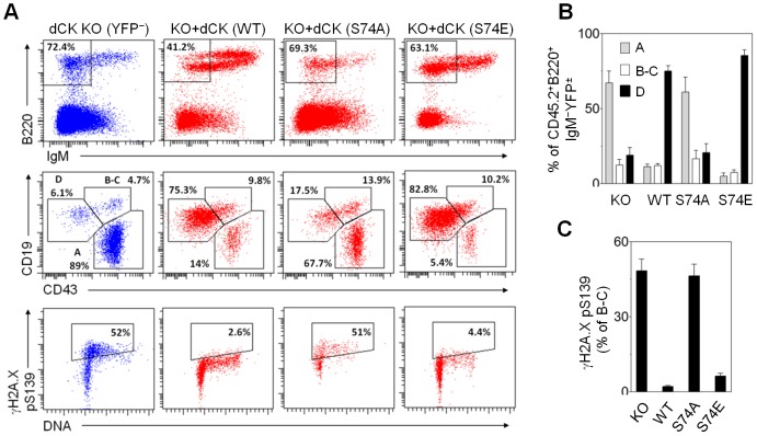
Efficient and adequate generation of deoxyribonucleotides is critical to successful DNA repair. We show that ataxia telangiectasia mutated (ATM) integrates the DNA damage response with DNA metabolism by regulating the salvage of deoxyribonucleosides. Specifically, ATM phosphorylates and activates deoxycytidine kinase (dCK) at serine 74 in response to ionizing radiation (IR). Activation of dCK shifts its substrate specificity toward deoxycytidine, increases intracellular dCTP pools post IR, and enhances the rate of DNA repair. Mutation of a single serine 74 residue has profound effects on murine T and B lymphocyte development, suggesting that post-translational regulation of dCK may be important in maintaining genomic stability during hematopoiesis. Using [(18)F]-FAC, a dCK-specific positron emission tomography (PET) probe, we visualized and quantified dCK activation in tumor xenografts after IR, indicating that dCK activation could serve as a biomarker for ATM function and DNA damage response in vivo. In addition, dCK-deficient leukemia cell lines and murine embryonic fibroblasts exhibited increased sensitivity to IR, indicating that pharmacologic inhibition of dCK may be an effective radiosensitization strategy.
Conflict of interest statement
Figures


Similar articles
-
A crucial role for ATR in the regulation of deoxycytidine kinase activity.Biochem Pharmacol. 2016 Jan 15;100:40-50. doi: 10.1016/j.bcp.2015.11.022. Epub 2015 Nov 24. Biochem Pharmacol. 2016. PMID: 26620371
-
Deoxycytidine kinase regulates the G2/M checkpoint through interaction with cyclin-dependent kinase 1 in response to DNA damage.Nucleic Acids Res. 2012 Oct;40(19):9621-32. doi: 10.1093/nar/gks707. Epub 2012 Jul 31. Nucleic Acids Res. 2012. PMID: 22850745 Free PMC article.
-
The Role of Deoxycytidine Kinase (dCK) in Radiation-Induced Cell Death.Int J Mol Sci. 2016 Nov 21;17(11):1939. doi: 10.3390/ijms17111939. Int J Mol Sci. 2016. PMID: 27879648 Free PMC article.
-
New insights into the synergism of nucleoside analogs with radiotherapy.Radiat Oncol. 2013 Sep 26;8:223. doi: 10.1186/1748-717X-8-223. Radiat Oncol. 2013. PMID: 24066967 Free PMC article. Review.
-
ATM: the protein encoded by the gene mutated in the radiosensitive syndrome ataxia-telangiectasia.Int J Radiat Biol. 1999 Oct;75(10):1201-14. doi: 10.1080/095530099139359. Int J Radiat Biol. 1999. PMID: 10549596 Review.
Cited by
-
Nucleotide salvage deficiencies, DNA damage and neurodegeneration.Int J Mol Sci. 2015 Apr 27;16(5):9431-49. doi: 10.3390/ijms16059431. Int J Mol Sci. 2015. PMID: 25923076 Free PMC article. Review.
-
Human Biodistribution and Radiation Dosimetry of 18F-Clofarabine, a PET Probe Targeting the Deoxyribonucleoside Salvage Pathway.J Nucl Med. 2017 Mar;58(3):374-378. doi: 10.2967/jnumed.116.182394. Epub 2016 Nov 3. J Nucl Med. 2017. PMID: 27811125 Free PMC article.
-
dCK negatively regulates the NRF2/ARE axis and ROS production in pancreatic cancer.Cell Prolif. 2018 Aug;51(4):e12456. doi: 10.1111/cpr.12456. Epub 2018 Apr 27. Cell Prolif. 2018. PMID: 29701272 Free PMC article.
-
[18F]CFA as a clinically translatable probe for PET imaging of deoxycytidine kinase activity.Proc Natl Acad Sci U S A. 2016 Apr 12;113(15):4027-32. doi: 10.1073/pnas.1524212113. Epub 2016 Mar 28. Proc Natl Acad Sci U S A. 2016. PMID: 27035974 Free PMC article.
-
Role of Damage DNA-Binding Protein 1 in Pancreatic Cancer Progression and Chemoresistance.Cancers (Basel). 2019 Dec 12;11(12):1998. doi: 10.3390/cancers11121998. Cancers (Basel). 2019. PMID: 31842285 Free PMC article.
References
-
- Mathews CK (2006) DNA precursor metabolism and genomic stability. Faseb J 20: 1300–1314. - PubMed
-
- Reichard P (1988) Interactions between deoxyribonucleotide and DNA synthesis. Annu Rev Biochem 57: 349–374. - PubMed
-
- Sabini E, Ort S, Monnerjahn C, Konrad M, Lavie A (2003) Structure of human dCK suggests strategies to improve anticancer and antiviral therapy. Nat Struct Biol 10: 513–519. - PubMed
-
- Csapo Z, Keszler G, Sasvari-Szekely M, Smid K, Noordhuis P, et al. (1998) Similar changes were induced by cladribine and by gemcitabine, in the deoxypyrimidine salvage, during short term treatments. Adv Exp Med Biol 431: 525–529. - PubMed
Publication types
MeSH terms
Substances
Grants and funding
LinkOut - more resources
Full Text Sources
Other Literature Sources
Molecular Biology Databases
Research Materials
Miscellaneous
