. 2014 Aug 7;10(8):e1004514.
doi: 10.1371/journal.pgen.1004514. eCollection 2014 Aug.

Syndecan-1 is required to maintain intradermal fat and prevent cold stress

Affiliations

Syndecan-1 is required to maintain intradermal fat and prevent cold stress

Ildiko Kasza et al. PLoS Genet. .

Abstract

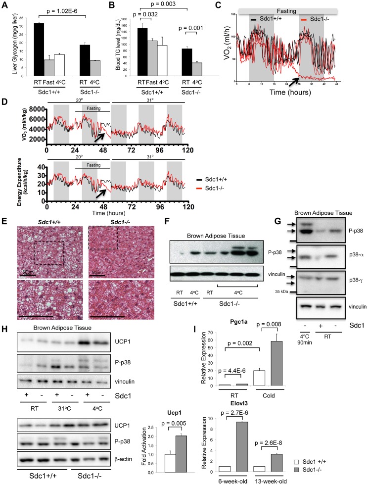
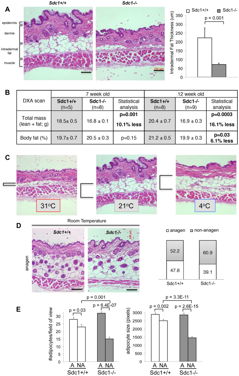
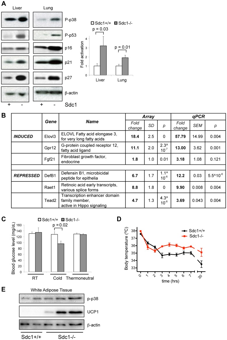
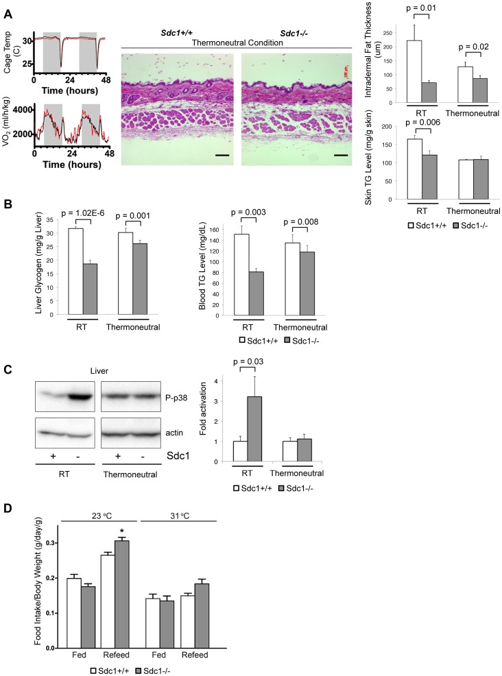
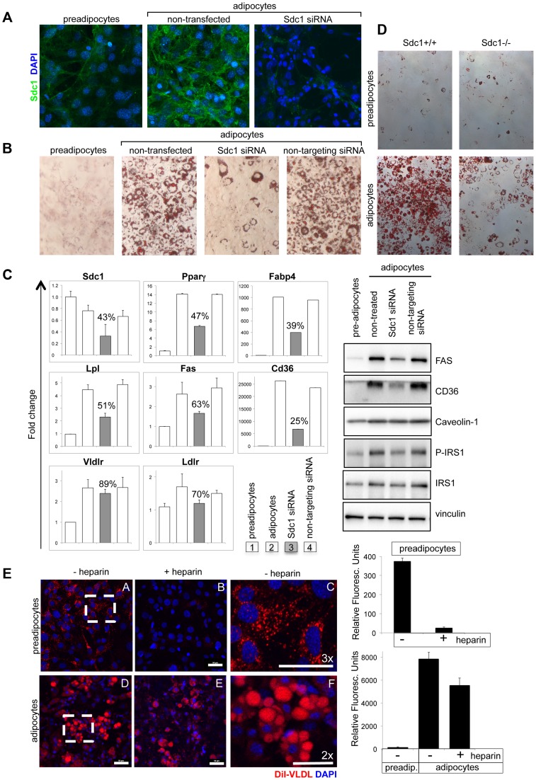
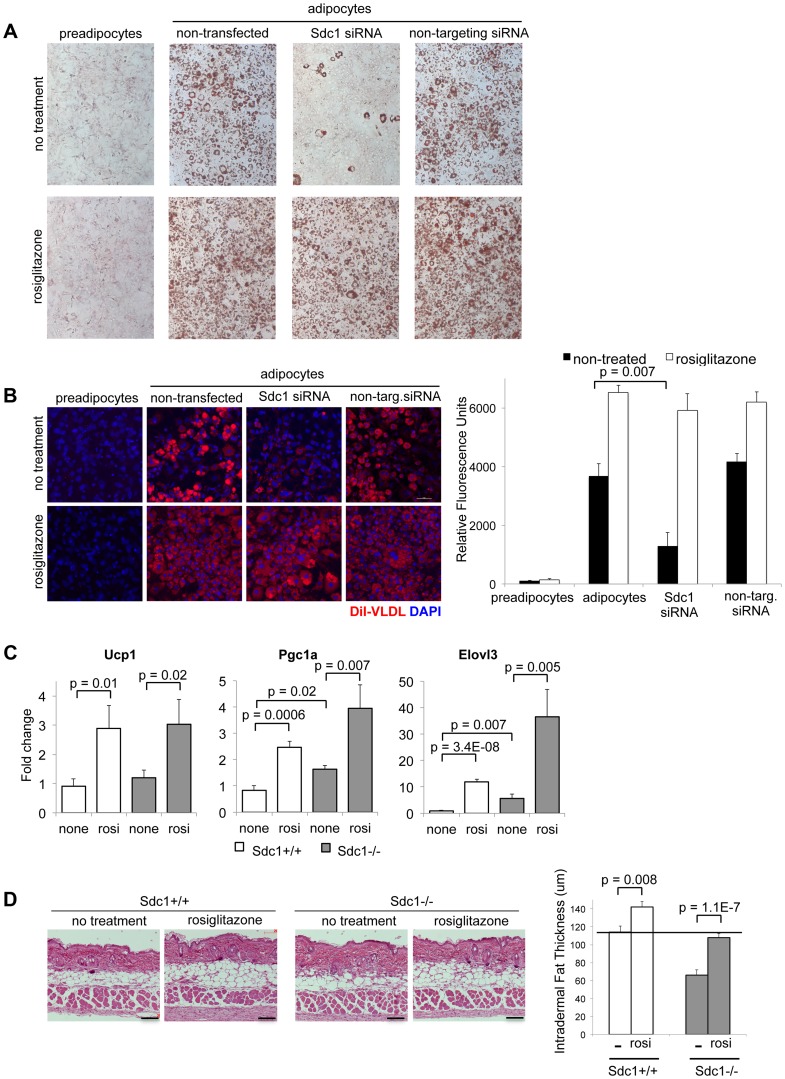
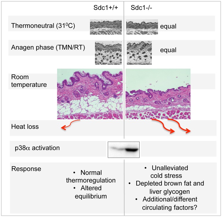
Homeostatic temperature regulation is fundamental to mammalian physiology and is controlled by acute and chronic responses of local, endocrine and nervous regulators. Here, we report that loss of the heparan sulfate proteoglycan, syndecan-1, causes a profoundly depleted intradermal fat layer, which provides crucial thermogenic insulation for mammals. Mice without syndecan-1 enter torpor upon fasting and show multiple indicators of cold stress, including activation of the stress checkpoint p38α in brown adipose tissue, liver and lung. The metabolic phenotype in mutant mice, including reduced liver glycogen, is rescued by housing at thermoneutrality, suggesting that reduced insulation in cool temperatures underlies the observed phenotypes. We find that syndecan-1, which functions as a facultative lipoprotein uptake receptor, is required for adipocyte differentiation in vitro. Intradermal fat shows highly dynamic differentiation, continuously expanding and involuting in response to hair cycle and ambient temperature. This physiology probably confers a unique role for Sdc1 in this adipocyte sub-type. The PPARγ agonist rosiglitazone rescues Sdc1-/- intradermal adipose tissue, placing PPARγ downstream of Sdc1 in triggering adipocyte differentiation. Our study indicates that disruption of intradermal adipose tissue development results in cold stress and complex metabolic pathology.


Conflict of interest statement

The authors have declared that no competing interests exist.

Figures

Figure 1
Figure 1. Sdc1−/− mice are chronically cold- stressed.
A. Liver glycogen was measured for Sdc1−/− and wild type BALB/c mice under normal housing conditions (room temperature; RT; 21°C), after a 24-hour fast or after acute cold stress (90 mins at 4°C; n = 6). B. Serum triglycerides were measured for the same conditions. C. Mice of both genotypes were housed in metabolic cages at 23°C, fasted and their metabolic rates were measured (oxygen consumption, expressed as VO2). In this example, Sdc1−/− mice entered torpor after 24 hours (indicated as a precipitous drop in O2 consumption for two out of four Sdc1−/− mice in this experiment). None of the wild type (0/4) mice triggered torpor. D. In this example, measurements from 4 BALB/c (black lines) and 4 Sdc1−/− (red lines) mice were averaged (3/4 Sdc1−/− mice and 0/4 BALB/c mice entered torpor, indicated by the blue arrows). The housing temperature was shifted up to 31°C to illustrate their complete recovery (shown as VO2/oxygen consumption and energy expenditure curves over 3 days). E. Brown adipose tissues were harvested from Sdc1−/− and BALB/c mice housed at 21°C. H&E-stained paraffin sections show that Sdc1−/− brown adipose shows depleted lipid stores (white cytoplasmic inclusions). F. To test for cold stress, lysates of brown adipose tissue (shown here from three independent mice) were assayed for activation of the checkpoint, pT180Y182 p38α, either at room temperature or after acute cold stress (4°C). G. To confirm which isotypes of p38 were present and activated, the pattern of phospho-p38 was compared to Western blots probed with isotype-specific antibodies. H. Lysates were probed for UCP1, a marker of uncoupling in brown adipose tissue. Top panel, BAT from mice housed at various housing temperatures; bottom panel, assay of relative levels of UCP1 in Sdc1−/− mice housed at RT. I. To test whether the BAT of Sdc1−/− mice had initiated the full thermogenic program, levels of cold stress-induced mRNAs were measured by qPCR for tissues isolated from mice housed at RT and 4°C/cold (5 days) (PGC1α, n = 3) and for mice of different ages (6- and 13-week-old; Elovl3).
Figure 2
Figure 2. Sdc1−/− mice have thinner intradermal fat.
A. Paraffin-embedded belly skin samples of Sdc1−/− and wild type BALB/c mice housed at 23°C were sectioned, H&E stained and the thickness of intradermal fat was quantified for non-anagen stage skin samples, measuring from muscle to dermis (n = 18 and 21 respectively). B. Body mass index was measured by DXA imaging for two groups of Sdc1−/− and wild type mice (as indicated). C. Paraffin-embedded belly skin samples of wild type BALB/c mice, housed at 31°C (5 days), 23°C (constant housing) and 4°C (5 days), were sectioned to illustrate the effect of housing temperature on intradermal fat deposits. Intradermal fat is indicated by brackets. (Back skin showed similar effects, but all skins compared for any one experiment come from the same location). D. Samples of anagen-stage belly skin for Sdc1−/− and wild type BALB/c mice housed at 23°C were stained with H&E. The hair follicle cycle is asynchronous in adults; anagen-stage is defined by the intrusion of hair follicles to the bottom of the intradermal fat layer, and a high epithelial mitotic index (Fig. S3). Skin samples were scored as anagen or non-anagen for cohorts of Sdc1−/− and wild type BALB/c mice, and the proportion of each is illustrated (right hand side), to provide an estimate of relative frequency of anagen-stage skin. E. Morphometric analysis of adipocytes shows that size and number of adipocytes is normal in Sdc1−/− skin during anagen, but both are reduced in non-anagen stage Sdc1−/− skins (n = 6).
Figure 3
Figure 3. Sdc1−/− mice show systemic hyper-activation of p38α, and other metabolic markers of cold stress.
A. Tissue lysates from livers and lungs of Sdc1−/− and wild type BALB/c mice under normal housing conditions were analyzed for activation of p38 (P-p38), and the effectors downstream of p38 signaling. Western blots were quantified to show relative p38α activation. B. Transcriptome analysis of liver mRNA showed some specific changes in Sdc1−/− livers. C. Sdc1−/− mice showed normal blood glucose levels until mice were cold-stressed (4°C, 90 mins) (n = 4). D. Body temperature was normal in Sdc1−/− mice until mice were cold-stressed (n = 7). E. Tissue lysates from peri-gonadal white adipose tissues (WAT) were analyzed for expression of UCP1, as a measure of cold activation and browning.
Figure 4
Figure 4. Thermoneutral housing rescues the Sdc1−/− phenotype.
A. To illustrate the energy required to respond to a transition between 31°C (thermoneutral temperature) and 23°C, the oxygen uptake of mice in metabolic cages was measured (black, BALB/c; red, Sdc1−/−). Skin samples from mice housed at 31°C for 2 weeks were paraffin-embedded, sectioned, and H&E stained. Results showed that intradermal fat was thinner and almost equal for both BALB/c and Sdc1−/− mice (quantified at right hand side; n = 6). Skin triglycerides (TG) were also measured (triglyceride derives both from intradermal fat and from the epidermal layer itself, including the sebaceous glands). B. Liver glycogen and blood glucose levels are shown for mice housed at 31°C (and 23°C, RT), showing almost complete rescue. C. Tissue lysates from livers of mice housed at thermoneutrality showed that p38 activation in Sdc1−/− mice was equal to wild type BALB/c mice. D. Quantitation of relative food intake (fed, or fasted for 48 hours and re-fed) (n = 8 for each of Sdc1−/− and BALB/c wild type mice), to show that Sdc1−/− mice are hyperphagic during recovery from fasting.
Figure 5
Figure 5. Loss of Sdc1 ablates adipocyte differentiation in vitro.
A. 3T3-L1 cells (pre-adipocytes), 3T3-L1 cells 8 days after initiation of the differentiation protocol (adipocytes) and 3T3-L1 cells after knockdown of Sdc1 with siRNA were assayed for Sdc1 expression by immunofluorescent antibody staining. B. A similar series of cultured cells were stained with Oil Red-O, a dye that dissolves in lipid drops accumulating in differentiated adipocytes. C. Cell lysates were assayed for markers of differentiation by qPCR (PPARγ, peroxisome proliferator-activated receptor gamma; FABP4, fatty acid binding protein-4; LPL, lipoprotein lipase; FASN, fatty acid synthase; CD36, thrombospondin receptor) and by Western blotting (FasN, Cd36, activated phospho-IRS1). D. Ear mesenchymal stem cells (eMSCs) were isolated from Sdc1−/− and BALB/c mice, transferred to culture, and induced to differentiate. Differentiation was visualized by Oil Red-O staining. Corresponding nuclear stains (Fig. S6C) illustrate that the cell densities are approximately similar. E. To evaluate the impact of a heparan sulfate mimetic, heparin, on the ability of 3T3-L1 preadipocytes (A,B,C) and adipocytes (D,E,F) to take up VLDL, cells were incubated in presence (B,E) or absence (A,C,D,F) of 200 µM heparin with di-I labeled VLDL for 3 hours at 37°C. Fields from A and D were magnified (C and F respectively) to show the size and morphology of the red-stained vesicles. Uptake was quantified (right hand side panel). Lower concentrations of heparin showed similar effects (50 µM; data not shown).
Figure 6
Figure 6. Adipocyte differentiation can be rescued in vitro and in vivo by the PPARγ agonist, rosiglitazone.
A. 3T3-L1 cells, treated as indicated, were stained for Oil Red O to assess their differentiation. Rosiglitazone (2 µM) was added with the differentiation medium for 2 days. B. For similar cultures, VLDL uptake was measured. C. Rosiglitazone (0.015% diet for 5 days) was administered to Sdc1−/− and wild type mice, and reporters of pPARγ activity (mRNAs for Ucp1, Pgc1α and Elovl3) were measured by qPCR in mRNA extracts of white adipose tissue. D. Skins from rosiglitazone- and control-diet fed mice were paraffin embedded and sectioned to determine the thickness of intradermal fat (n = 8).
Figure 7
Figure 7. Summary scheme of the effects of deficient intradermal fat in Sdc1−/− mice.
The thickness of intradermal fat in non-anagen phases is set by ambient temperature, and is 80% depleted in Sdc1−/− mice housed at room temperatures. During anagen phase (when intradermal fat expands in response to local cues), the thickness of Sdc1−/− intradermal fat is high and normal; in warm temperatures, the intradermal fat of Sdc1−/− mice is thin and normal. Heat loss from skin containing 40 µM intradermal fat is calculated to be at least 2-fold higher than skin with 200 µM of intradermal fat. This heat loss leads to systemic p38α activation throughout intra-abdominal tissues, and a condition of “unalleviated cold stress” in Sdc1−/− mice.

References

    1. Karp CL (2012) Unstressing intemperate models: how cold stress undermines mouse modeling. The Journal of experimental medicine 209: 1069–1074. - PMC - PubMed
    1. Golozoubova V, Gullberg H, Matthias A, Cannon B, Vennstrom B, et al. (2004) Depressed thermogenesis but competent brown adipose tissue recruitment in mice devoid of all hormone-binding thyroid hormone receptors. Molecular endocrinology 18: 384–401. - PubMed
    1. Overton JM, Williams TD (2004) Behavioral and physiologic responses to caloric restriction in mice. Physiology & behavior 81: 749–754. - PubMed
    1. Gordon CJ (2012) Thermal physiology of laboratory mice: Defining thermoneutrality. Journal of Thermal Biology 37: 654–685.
    1. Nguyen KD, Qiu Y, Cui X, Goh YP, Mwangi J, et al. (2011) Alternatively activated macrophages produce catecholamines to sustain adaptive thermogenesis. Nature 480: 104–108. - PMC - PubMed

Publication types

MeSH terms

LinkOut - more resources