. 2014 Aug 7;10(8):e1004540.
doi: 10.1371/journal.pgen.1004540. eCollection 2014 Aug.

The cohesin subunit Rad21 is required for synaptonemal complex maintenance, but not sister chromatid cohesion, during Drosophila female meiosis

Affiliations

The cohesin subunit Rad21 is required for synaptonemal complex maintenance, but not sister chromatid cohesion, during Drosophila female meiosis

Evelin Urban et al. PLoS Genet. .

Abstract

Replicated sister chromatids are held in close association from the time of their synthesis until their separation during the next mitosis. This association is mediated by the ring-shaped cohesin complex that appears to embrace the sister chromatids. Upon proteolytic cleavage of the α-kleisin cohesin subunit at the metaphase-to-anaphase transition by separase, sister chromatids are separated and segregated onto the daughter nuclei. The more complex segregation of chromosomes during meiosis is thought to depend on the replacement of the mitotic α-kleisin cohesin subunit Rad21/Scc1/Mcd1 by the meiotic paralog Rec8. In Drosophila, however, no clear Rec8 homolog has been identified so far. Therefore, we have analyzed the role of the mitotic Drosophila α-kleisin Rad21 during female meiosis. Inactivation of an engineered Rad21 variant by premature, ectopic cleavage during oogenesis results not only in loss of cohesin from meiotic chromatin, but also in precocious disassembly of the synaptonemal complex (SC). We demonstrate that the lateral SC component C(2)M can interact directly with Rad21, potentially explaining why Rad21 is required for SC maintenance. Intriguingly, the experimentally induced premature Rad21 elimination, as well as the expression of a Rad21 variant with destroyed separase consensus cleavage sites, do not interfere with chromosome segregation during meiosis, while successful mitotic divisions are completely prevented. Thus, chromatid cohesion during female meiosis does not depend on Rad21-containing cohesin.


Conflict of interest statement

The authors have declared that no competing interests exist.

Figures
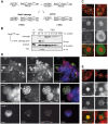
Figure 1
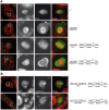
Figure 1. Rad21TEV-myc cleavage by TEV protease expression during oogenesis results in cohesin dissociation from chromatin.
(A) Crossing scheme illustrating the generation of females, in which the solely expressed Rad21 variant Rad21TEV-myc is cleaved during oogenesis due to Gal4 mediated expression of TEV protease (+TEV) as well as of control sibling females (−TEV). Rad21ex, deletion allele of Rad21. (B) Extracts were prepared from stage 14 oocytes obtained from control females (w1, or −TEV females) or from females expressing TEV protease in the Rad21ex, Rad21TEV-myc homozygous background (+TEV). Proteins were separated by PAGE, blotted and the blot was probed with antibodies against the myc-epitope (top panel), against α-tubulin as loading control (middle panel), and against the V5 epitope to monitor TEV protease expression (bottom panel). The numbers of oocyte equivalents are given on top of the lanes. (C) Immunofluorescence analysis of stage 4–5 egg chambers from Rad21 mutant females (+TEV) or sibling females (−TEV). DNA was stained with Hoechst 33258 and Rad21TEV-myc was labeled with anti-myc antibodies. In the upper row, an overview of the egg chambers is presented and the oocyte nucleus is shown enlarged in the other panels. In the merged images, DNA is shown in red and the myc-signal in green. (D) Chromosome spread analysis of germaria from females expressing Rad21TEV-myc. Within the partially dissociated germarium, some nuclei show the thread-like pattern of C(3)G staining typical for the synaptonemal complex (filled arrowheads in the top panel). In the same nuclei, myc signals are also thread-like and in nuclei of pro-nurse cells, which are negative for C(3)G staining, diffuse myc staining indicates Rad21TEV-myc association throughout chromatin (open arrowhead in the enlargements in the bottom panel). In the merged images, DNA is shown in blue, anti-myc in red and C(3)G in green. (E) Immunofluorescence analysis of stage 4–5 egg chambers from Rad21 mutant females (+TEV) or sibling females (−TEV). DNA was stained with Hoechst 33258 and SMC1 with anti-SMC1 antibodies. In the upper row, an overview of the egg chambers is presented and the oocyte nucleus is shown enlarged in the other panels. In the merged images, DNA is shown in red and the SMC1-signal in green. Images are single confocal sections. Exposure times and processing were identical for the images +/− TEV. Scale bars are 5 µm.
Figure 2
Figure 2. Premature Rad21TEV-myc cleavage during oogenesis results in precocious SC disassembly.
(A) Immunofluorescence analysis of stage 4–5 egg chambers from wild type females (w1), females with GAL4-driven expression of TEV protease in a Rad21 wild type background (mat-Gal4/UAS-TEV), females expressing only GAL4 in a Rad21TEV-myc rescue background (mat-Gal4/CyO; Rad21ex, Rad21TEV-myc/Rad21ex, Rad21TEV-myc), or females with GAL4-driven expression of TEV protease in a Rad21TEV-myc rescue background (mat-Gal4/UAS-TEV; Rad21ex, Rad21TEV-myc/Rad21ex, Rad21TEV-myc). DNA was stained with Hoechst 33258 and C(3)G was labeled with anti-C(3)G antibodies. In the left column, an overview of the egg chambers is presented and the oocyte nucleus is shown enlarged in the other panels. In the merged images, DNA is shown in red and the C(3)G-signal in green. Note the enrichment of C(3)G signal in the nucleoplasm after TEV-mediated Rad21TEV-myc cleavage (bottom panels). (B) Egg chambers from females expressing C(2)M-HA under control of the c(2)M genomic regulatory sequences in a Rad21 mutant background (UAS-TEV, C(2)M-HA/mat-Gal4; Rad21ex, Rad21TEV-myc/Rad21ex, Rad21TEV-myc) or from sibling females not expressing TEV protease (UAS-TEV, C(2)M-HA/CyO; Rad21ex, Rad21TEV-myc/Rad21ex, Rad21TEV-myc) were analyzed by immunolabelling with anti-HA antibodies. Images are single confocal sections. Exposure times and processing were identical for the images +/− TEV. Scale bars are 5 µm.
Figure 3
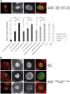
Figure 3. Premature SC disassembly can be triggered by Rad21 removal using different driver/transgene combinations.
(A) Immunofluorescence analysis of a stage 4–5 egg chamber from a female expressing TEV protease driven by nos-GAL4 in a Rad21TEV-myc rescue background. DNA was stained with Hoechst 33258 and C(3)G was labeled with anti-C(3)G antibodies. In the merged images, DNA is shown in red and the C(3)G-signal in green. The quantification illustrates the mean stage of SC disassembly in ovarioles of females with the indicated genotype. +TEV, TEV protease expression driven by nos-GAL4; −TEV, sibling controls not expressing TEV protease; Rad21TEV, indicates presence of the recombinant chromosome Rad21ex, Rad21TEV-myc. Rad21-EGFP, indicates presence of the recombinant chromosome Rad21ex, Rad21-EGFP; +deGradFP; NSlmb-vhhGFP4 expression driven by nos-GAL4; -deGradFP, sibling controls not expressing NSlmb-vhhGFP4. Black bars, TEV cleavage site position at aa 271 of Rad21; dark gray bars, TEV cleavage site position at aa 550 of Rad21; light grey bars, presence of the recombinant chromosome Rad21ex, Rad21-EGFP; white bars, controls expressing TEV protease in a wild type background (+TEV, +/+) or Rad21ex3 heterozygous females not expressing any transgene (Rad21*/+). In each case, 33 to 34 ovarioles were scored, except for +deGradFP, Rad21-EGFP/+ (21 ovarioles). Error bars represent standard error. ***: p<0.0001; **: p = 0.0002; as determined by pairwise comparisons using the Mann-Whitney U-test. (B) Immunofluorescence analysis of stage 4–5 egg chambers from females expressing TEV protease driven by mat-GAL4 in a Rad21ex, Rad21TEV-myc heterozygous background (mat-Gal4/UAS-TEV; Rad21ex, Rad21TEV-myc/TM3, Sb) or control females heterozygous for the Rad21 excision allele (Rad21ex/TM3, Sb). DNA was stained with Hoechst 33258 and C(3)G or SMC1 were labelled with specific antibodies. In the left column, an overview of the egg chambers is presented and the oocyte nucleus is shown enlarged in the other panels. In the merged images, DNA is shown in red and the C(3)G-signal/SMC1-signal in green. Note that even in the presence of one wild type Rad21 allele, cohesin leaves chromatin and the SC disassembles prematurely after forced Rad21 cleavage. Scale bars are 5 µm.
Figure 4
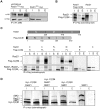
Figure 4. C(2)M physically interacts with Rad21.
(A) Extracts from 0–1.5 h old embryos expressing either h old embryos expressing either gC(2)M-HA together with Rad21TEV-myc or just Rad21TEV-myc were subjected to immunoprecipitation (IP) with mouse anti-HA antibodies. Bound proteins were eluted (E), separated by SDS-PAGE together with input (I) and supernatant after IP (S) samples, and analyzed by western blotting (WB). The blotted samples were probed with anti-HA antibodies to control for immunoprecipitation efficiency and anti-myc antibodies to assess co-precipitation of Rad21TEV-myc. The samples were run on the same gel but not immediately adjacent to each other. Lanes removed from the image are indicated by the vertical black line (B) Full length versions of Rad21 and Flag-epitope tagged C(2)M were synthesized by coupled in vitro transcription/translation (IVT) in the presence of [35S]methionine. IVT reactions were subjected to immunoprecipitation using anti-Flag antibodies. Radioactively labelled proteins were detected by autoradiography. The samples were run on the same gel but not immediately adjacent to each other. Lanes removed from the image are indicated by the vertical black line (C) Schematic of the various Rad21 and Flag-C(2)M fragments assayed for interaction in the coupled IVT-IP experiments. Rad21 fragments were untagged, while all C(2)M fragments were N-terminally fused to 3 copies of the Flag epitope. The proteins were either of full length (FL) or represented the N-terminal part (N), the middle part (M) or the C-terminal part (C) of Rad21 or C(2)M. After IVT-IP using anti-Flag antibodies the samples (I, input; S, supernatant, E, eluate) were separated by SDS-PAGE and radioactively labelled proteins were detected by autoradiography. The migration position of the various fragments is indicated on the left. (D) Coupled IVT-IP of full-length versions of SMC1, Rad21, and myc-C(2)M. After IP using anti-myc antibodies, input (I) and eluate (E) fractions were analyzed. Note that IVT of SMC1 resulted in two protein species, as indicated by asterisks on the right.
Figure 5
Figure 5. Ectopic Rad21 cleavage does not result in metaphase I alignment defects.
(A) Schematic illustrating the FISH probes used to detect the X and 4th chromosomes in late stage oocyte nuclei. Centromeres are indicated by dark grey circles. The X chromosome-specific 359 bp probe was labelled with Alexa 647 and the 4 bp probe was labelled with Alexa 647 and the 4th chromosome specific AATAT probe with Alexa 555. The images on the bottom show examples for the two different categories defined to score the FISH phenotype. The arrow indicates a supernumerary signal for the X chromosome-specific probe. Scale bar is 5 µm. (B) Quantification of the phenotypes of late stage oocyte nuclei after FISH using the X and 4th chromosome-specific probes. The females used to prepare the oocytes had the genotypes w1 (wt), or mat-GAL4/UAS-TEV; Rad21ex, Rad21TEV-myc/Rad21ex, Rad21TEV-myc (+TEV, Rad21TEV/Rad21TEV) or mat-GAL4/UAS-TEV; Rad21ex, Rad21TEV-myc/TM3, Sb (+TEV, Rad21TEV/+) or c(2)MEP2115/c(2)MEP2115 (c(2)M/c(2)M) or c(2)MEP2115, mat-GAL4/c(2)MEP2115, UAS-TEV; Rad21ex, Rad21TEV-myc/Rad21ex, Rad21TEV-myc (c(2)M/c(2)M, +TEV, Rad21TEV/Rad21TEV). The total numbers of oocytes scored are given on top of the diagram. (C) FISH analysis of anaphase II figures with probes detecting the X-chromosome (red in the merged images) and the 4th chromosome (green in the merged images) in eggs laid by females with the genotype mat-GAL4/UAS-TEV; Rad21ex, Rad21TEV-myc/Rad21ex, Rad21TEV-myc. In 82/83 cases, a normal 1∶1∶1∶1 distribution was observed for both probes (left panels). In 1/83 cases, a 0∶0∶2∶2 distribution for the X-chromosome was recorded, indicative of non-disjunction in meiosis I (right panels).
Figure 6
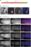
Figure 6. Expression of Rad21 with mutated separase cleavage sites does not impair meiotic divisions.
(A) Schematic illustration of the Rad21 variant with mutated separase cleavage sites (Rad21NC-myc). The arginines within the separase consensus sites at positions 175 and 474 were changed to alanines. (B) Chromosome spread analysis of germaria from females expressing Rad21NC-myc under control of MTD-GAL4. The non-cleavable Rad21 variant co-localizes with the synaptonemal complex component C(3)G indicating the incorporation of Rad21NC-myc into meiotic chromatin. In the merged image, DNA is shown in blue, the myc-signal in red, and the C(3)G-signal in green. Scale bar is 5 µm. (C) Embryos from mothers expressing Rad21NC-myc under control of MTD-GAL4 showed normal meiosis II figures (upper row). Each of the four meiotic products contains one X-chromosome-specific FISH signal. During later stages, three meiotic products collapse into the polar body, containing three X-chromosome-specific FISH signals (second row; inset). The zygotic nucleus appears hypercondensed. In the rare cases where multiple DNA masses were apparent within the embryo, they frequently exhibited pronounced anaphase bridges (defective mitoses; last row). Despite these defects the polar bodies have normal appearance and exhibit three X-chromosome-specific FISH signals (second to last row). The images in the bottom two rows represent different focal planes of the same embryo. In the merged images, DNA is shown in blue and the FISH signal in red. Scale bar is 50 µm.

Similar articles

Cited by

References

    1. Petronczki M, Siomos MF, Nasmyth K (2003) Un menage a quatre: the molecular biology of chromosome segregation in meiosis. Cell 112: 423–440. - PubMed
    1. McKim KS, Green-Marroquin BL, Sekelsky JJ, Chin G, Steinberg C, et al. (1998) Meiotic synapsis in the absence of recombination. Science 279: 876–878. - PubMed
    1. Manheim EA, McKim KS (2003) The Synaptonemal complex component C(2)M regulates meiotic crossing over in Drosophila. Curr Biol 13: 276–285. - PubMed
    1. Page SL, Hawley RS (2001) c(3)G encodes a Drosophila synaptonemal complex protein. Genes Dev 15: 3130–3143. - PMC - PubMed
    1. Oliveira RA, Nasmyth K (2010) Getting through anaphase: splitting the sisters and beyond. Biochem Soc Trans 38: 1639–1644. - PubMed

Publication types

LinkOut - more resources