. 2014 Aug 7;9(8):e104544.
doi: 10.1371/journal.pone.0104544. eCollection 2014.

In vitro anti-influenza virus activities of a new lignan glycoside from the latex of Calotropis gigantea

Affiliations

In vitro anti-influenza virus activities of a new lignan glycoside from the latex of Calotropis gigantea

Supawadee Parhira et al. PLoS One. .

Abstract

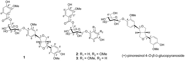
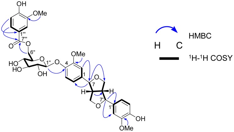
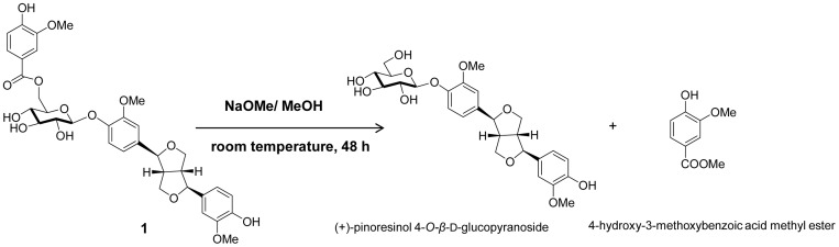
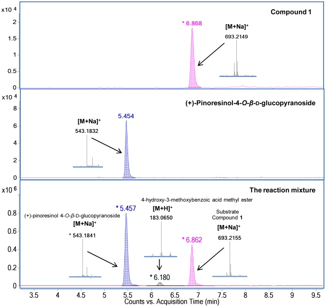
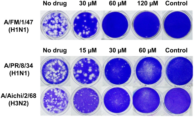
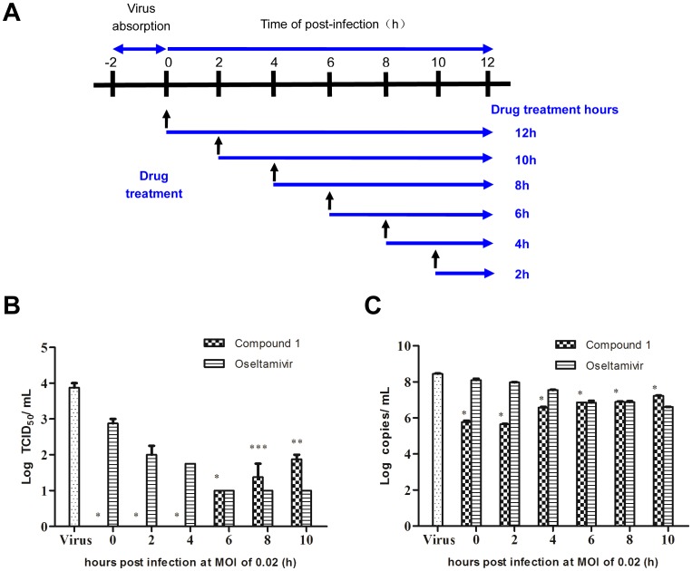
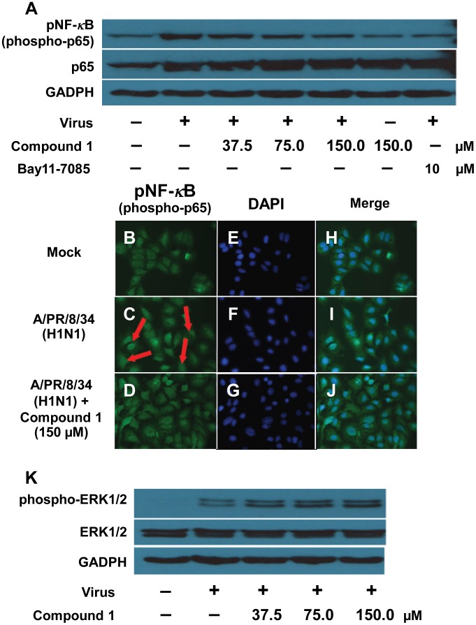
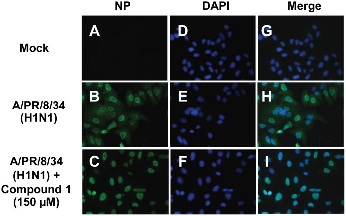
A new lignan glycoside, (+)-pinoresinol 4-O-[6″-O-vanilloyl]-β-D-glucopyranoside (1) and two known phenolic compounds, 6'-O-vanilloyltachioside (2) and 6'-O-vanilloylisotachioside (3) were isolated from the latex of Calotropis gigantea (Asclepiadaceae). The structure of the new compound was elucidated by using spectroscopic and chemical methods. Three isolates (1-3) and one authentic compound, (+)-pinoresinol 4-O-β-D-glucopyranoside, were screened for A/PR/8/34 (H1N1) inhibitory activity by cytopathic effect (CPE) inhibition assay on MDCK cells. Compound 1 showed inhibitory activity against A/PR/8/34 (H1N1). In sharp contrast, the other three compounds (2, 3 and (+)-pinoresinol 4-O-β-D-glucopyranoside) did not show such activity. An analysis of structure-activity relationship between 1 and (+)-pinoresinol 4-O-β-D-glucopyranoside revealed that the presence of a vanilloyl group in the sugar moiety of 1 is crucial for its anti-influenza virus activity. Compound 1 was further evaluated for in vitro inhibitory activities against a panel of human and avian influenza viruses by CPE inhibition assay. It showed inhibitory effect against human influenza viruses in both subtypes A and B (IC50 values around 13.4-39.8 µM with SI values of 3.7-11.4), while had no effect on avian influenza viruses. Its antiviral activity against human influenza viruses subtype A was further confirmed by plaque reduction assay. The time course assay indicated that 1 exerts its antiviral activity at the early stage of viral replication. A mechanistic study showed that 1 efficiently inhibited influenza virus-induced activation of NF-κB pathway in a dose-dependent manner, but had no effect on virus-induced activation of Raf/MEK/ERK pathway. Further studies demonstrated that nuclear translocation of transcription factor NF-κB induced by influenza virus was significantly blocked by 1, meanwhile, nuclear export of viral ribonucleoproteins was also effectively inhibited. These findings suggest that this new lignan glycoside from Calotropis gigantea, may have therapeutic potential in influenza virus infection through inhibition of NF-κB pathway and viral ribonucleoproteins nuclear export.


Conflict of interest statement

Competing Interests: The authors have declared that no competing interests exist.

Figures

Figure 1
Figure 1. Chemical structures of compounds 1–3 and (+)-pinoresinol 4-O-β-d-glucopyranoside.
Figure 2
Figure 2. Selected HMBC and 1H–1H COSY correlations of compound 1.
Arrows indicate the heteronuclear multiple-bond correlation of hydrogen atom to neighboring carbon. Bold line demonstrated the correlations between two nearby hydrogen atoms.
Figure 3
Figure 3. Mild alkaline hydrolysis of compound 1.
Figure 4
Figure 4. UHPLC-MS chromatograms and HRESIMS data of the products from alkaline hydrolysis of compound 1.
The methanolic solution of compound 1 was treated with sodium methoxide to furnish (+)-pinoresinol 4-O-β-d-glucopyranoside and 4-hydroxy-3-methoxy benzoic acid methyl ester.
Figure 5
Figure 5. Plaque reduction assay of compound 1 against influenza viruses.
MDCK cells were infected with influenza viruses, including A/FM/1/47 (H1N1) (Top row), A/PR8/8/34 (H1N1) (Middle row) and A/Aichi2/68 (H3N2) (Bottom row) at 0.01 MOI for 2 h at 34°C. After viral adsorption, cell monolayer was covered with overlay medium containing compound 1 and further cultured at 34°C under 5% CO2 for 48 h. Then, the overlay medium was removed, and the cell monolayer was fixed with 10% formalin, stained with 1% crystal violet, and plaques were counted.
Figure 6
Figure 6. Time course assay of compound 1.
(A) An illustration scheme shows the time of addition of compound 1. (B–C) MDCK cells (2×10⁴ cells/well) in 48-well plates were infected with influenza virus A/PR/8/34 (H1N1) (MOI = 0.02). At post-infection, the medium was discarded and cells were washed with PBS three times. Compound 1 (134.3 µM) or oseltamivir (6.4 µM) was added at 0 h, 2 h, 4 h, 6 h, 8 h and 10 h after infection. At 12 h post-infection, the supernatants were collected and infectious titers were determined by CPE assay (B) and real time PCR assay (C). Data represent mean ± SD of 3 biological samples. Statistical significance was assessed by comparison between compound 1-treated group and virus control group by using student's t-test analysis (* p-value <0.001, ** p-value <0.01 and *** p-value <0.05).
Figure 7
Figure 7. Inhibition of influenza virus-induced NF-κB activation in A549 cells by compound 1.
Compound 1 inhibited the influenza virus-induced NF- κB activation (A–J) but did not affect virus-induced Raf/MEK/ERK pathway activation (K). (A) and (K): A549 cells were infected with A/PR/8/34 (H1N1) (MOI = 0.1) in the absence or presence of different concentrations of compound 1 or the specific NF-κB inhibitor Bay11-7085 (10 µM) alone. A549 cell lysates were subjected to Western blot with specific antibodies against phospho-NF-κB p65 (Ser536), total NF-κB p65, phosphorylated ERK1/2 and ERK1/2. Equal protein load was verified using pan-antisera to GADPH. (B–J): A549 cells were infected with A/PR/8/34 (H1N1) (MOI = 1) and stained for against phospho-NF-κB p65 (Ser536) at 10 h of post-infection (green). Cell nuclei were stained with DAPI (blue).
Figure 8
Figure 8. Effect of compound 1 on influenza virus-induced export of viral ribonucleoprotein complexes.
Immunofluorescence confocal microscopy was used to show the nuclear RNP export in the absence or presence of compound 1. A549 cells were infected with A/PR/8/34 (H1N1) (MOI = 1) and stained for influenza viral nucleoprotein (NP) at 10 h of post-infection (green). Cell nuclei were stained with DAPI (blue).

References

    1. World Health Organization (2014) Available: http://www.who.int/topics/influenza/en. Accessed: 2014 Apr 30.
    1. Gao HN, Lu HZ, Cao B, Du B, Shang H, et al. (2013) Clinical findings in 111 cases of influenza A (H7N9) virus infection. N Engl J Med 368: 2277–2285. - PubMed
    1. Gao GF (2014) Influenza and the live poultry trade. Science 344: 235. - PubMed
    1. Du J, Cross TA, Zhou HX (2012) Recent progress in structure-based anti-influenza drug design. Drug Discov Today 17: 1111–1120. - PMC - PubMed
    1. Deyde VM, Xu X, Bright RA, Shaw M, Smith CB, et al. (2007) Surveillance of resistance to adamantanes among influenza A(H3N2) and A(H1N1) viruses isolated worldwide. J Infect Dis 196: 249–257. - PubMed

Publication types

MeSH terms