. 2014 Aug 7;8(8):e2980.
doi: 10.1371/journal.pntd.0002980. eCollection 2014 Aug.

Neuropathogenesis of Japanese encephalitis in a primate model

Affiliations

Neuropathogenesis of Japanese encephalitis in a primate model

Khin Saw Aye Myint et al. PLoS Negl Trop Dis. .

Abstract

Background: Japanese encephalitis (JE) is a major cause of mortality and morbidity for which there is no treatment. In addition to direct viral cytopathology, the inflammatory response is postulated to contribute to the pathogenesis. Our goal was to determine the contribution of bystander effects and inflammatory mediators to neuronal cell death.

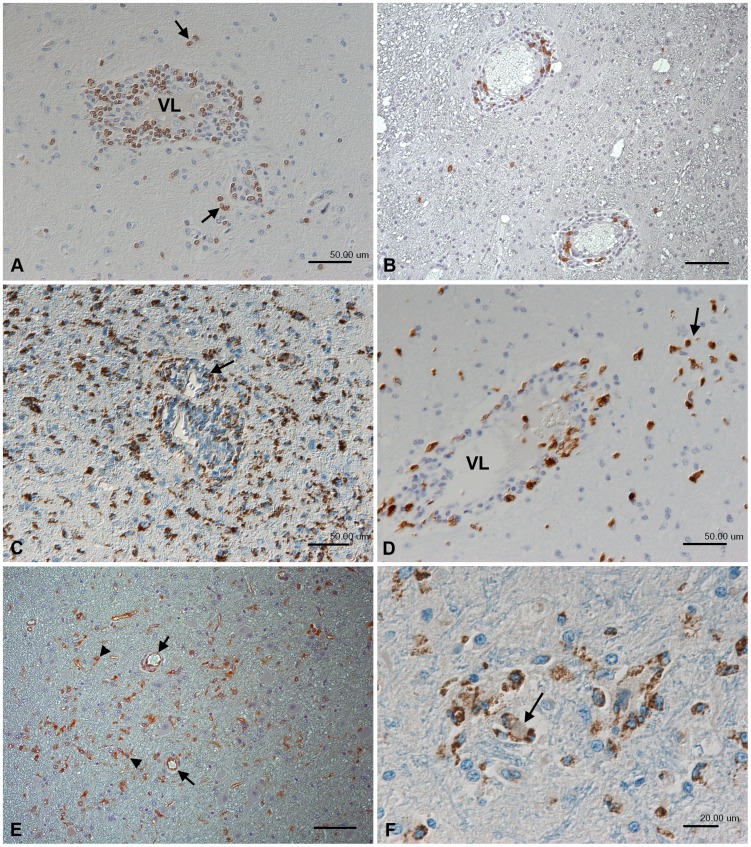
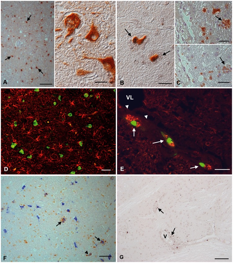
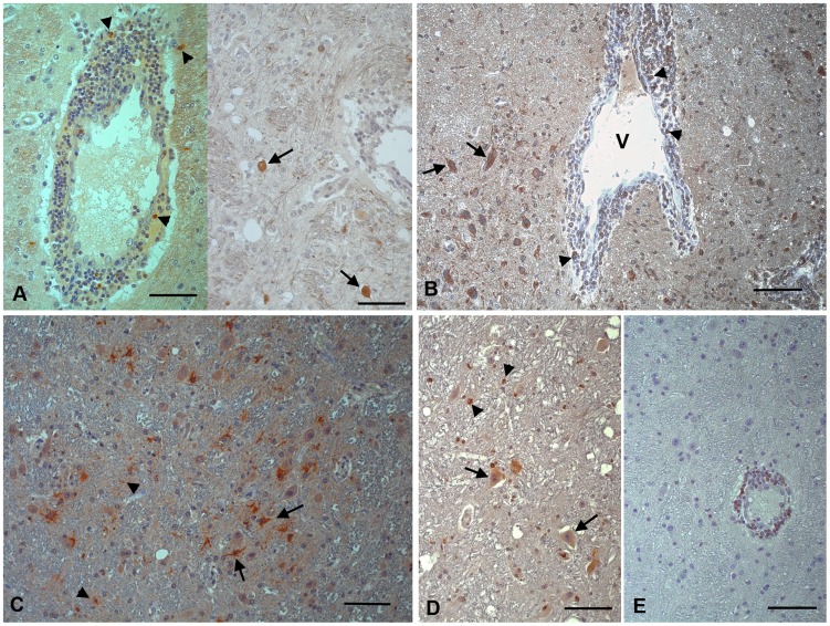
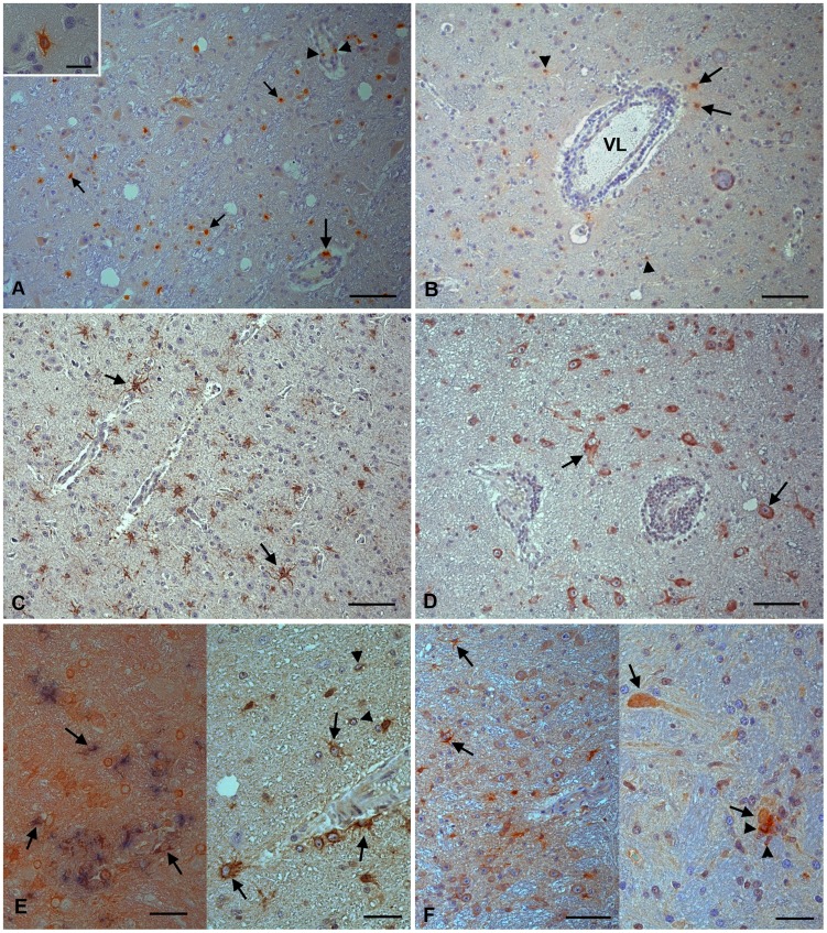
Methodology/principal findings: Material from a macaque model was used to characterize the inflammatory response and cytopathic effects of JE virus (JEV). Intranasal JEV infection induced a non-suppurative encephalitis, dominated by perivascular, infiltrates of mostly T cells, alongside endothelial cell activation, vascular damage and blood brain barrier (BBB) leakage; in the adjacent parenchyma there was macrophage infiltration, astrocyte and microglia activation. JEV antigen was mostly in neurons, but there was no correlation between intensity of viral infection and degree of inflammatory response. Apoptotic cell death occurred in both infected and non-infected neurons. Interferon-α, which is a microglial activator, was also expressed by both. Tumour Necrosis Factor-α, inducible nitric oxide synthase and nitrotyrosine were expressed by microglial cells, astrocytes and macrophages. The same cells expressed matrix metalloproteinase (MMP)-2 whilst MMP-9 was expressed by neurons.

Conclusions/significance: The results are consistent with JEV inducing neuronal apoptotic death and release of cytokines that initiate microglial activation and release of pro-inflammatory and apoptotic mediators with subsequent apoptotic death of both infected and uninfected neurons. Activation of astrocytes, microglial and endothelial cells likely contributes to inflammatory cell recruitment and BBB breakdown. It appears that neuronal apoptotic death and activation of microglial cells and astrocytes play a crucial role in the pathogenesis of JE.


Conflict of interest statement

The authors have declared that no competing interests exist.

Figures

Figure 1
Figure 1. Histopathological changes in the thalamus of a rhesus macaque (No. 2) after intranasal inoculation with JEV.
(A) Non-suppurative encephalitis, represented by moderate, lymphocyte-dominated perivascular infiltration. (B) Small vein with mild perivascular infiltration and activated endothelial cells (arrow). (C) The presence of serum, indicated by staining for von Willebrandt factor, in the parenchyma surrounding vessels with perivascular infiltrates (arrows) indicates marked vessel leakage. (D) Degenerating neuron (arrow) surrounded by glial cells (satellitosis). (E) Microglial nodule with occasional apoptotic cells (black arrow). (F) Staining for GFAP highlights the presence of large numbers of activated astrocytes (reactive astrocytosis). A, B, D, E: Hematoxylin-eosin stain. C, F: Indirect peroxidase method, NovaRed (C), DAB (F), hematoxylin counterstain. Scale bars: A, C, F = 50 µm; B, D, E = 20 µm.
Figure 2
Figure 2. Inflammatory response in the thalamus of rhesus macaques after intranasal inoculation with JEV ((No. 2 (A, B, E) and No. 9 (C, D, F)).
(A) CD3+ T cells dominate the perivascular infiltrates and are present in smaller numbers in the adjacent parenchyma (arrows). VL: vessel lumen. (B) CD20+ B cells represent a minority in the perivascular infiltrates. (C) Staining for CD68 identifies moderate numbers of macrophage/microglial cells within and surrounding the perivascular infiltrates (arrows) and highlights the large number of disseminated activated microglial cells in the adjacent parenchyma. (D) Macrophages in the perivascular infiltrates and the adjacent parenchyma (arrow) also express the myeloid/histiocyte antigen which indicates that they have recently been recruited from the blood. VL: vessel lumen. (E) Activated microglial cells also express major histocompatibility complex (MHC) class II antigen (arrowheads). MHC II is also expressed by vascular endothelial cells (arrows), confirming their activation. (F) Microglial nodule with central degenerate neuron (arrow), surrounded by CD68-positive microglial cells. Indirect peroxidise method, DAB, Papanicolaou's hematoxylin counterstain. Scale bars: A–E = 50 µm; F = 20 µm.
Figure 3
Figure 3. JEV target cells in the thalamus of rhesus macaques after intranasal inoculation with JEV ((No. 7 (A, B), No. 2 (C–G)).
(A) JEV antigen is seen in the majority of neurons (left: arrows). Right: Infected unaltered neurons express viral antigen in both cell body and cell processes. (B) JEV-infected neurons that are surrounded by microglial cells in satellitosis appear shrunken (arrows). (C) Microglial cells in particular in microglial nodules can be JEV-infected (top; arrow) and are identified based on their CD68 expression (bottom; arrow), as demonstrated in a consecutive section. (D) Dual staining for JEV antigen (FITC) and GFAP (Texas red) indicates that JEV does not infect astrocytes. (E) While endothelial cells (arrowheads) were not found to be JEV infected, perivascular macrophages in one animal were found to express JEV antigen (Texas Red); these cells were also undergoing apoptosis, since they were TUNEL-positive (FITC) (arrows). VL: vessel lumen. (F) Dual staining for JEV antigen (Vector Blue) and TUNEL (DAB) shows both the degenerating neurons and surrounding microglial cells in satellitosis undergo apoptosis (arrows). JEV-infected, apoptotic microglial cells (arrowhead) are also observed. (G) Occasional TUNEL-positive, apoptotic lymphocytes (arrows) are present in the perivascular infiltrates. V: vessel. Indirect peroxidase method (A–E, G), Vectastain Elite ABC-Alkaline Phosphatase Kit (F). DAB (A–G), BCIP/NBT blue (F), Papanicolaou's hematoxylin counterstain. Scale bars: A (left) = 100 µm; A (right), C = 25 µm; B, E = 20 µm; D, F, G = 50 µm.
Figure 4
Figure 4. Apoptosis related proteins in the thalamus of rhesus macaques after intranasal inoculation with JEV ((No. 2 (A, D, E), No. 9 (B), No. 11 (C)).
(A) Some leukocytes in the perivascular infiltrates (left, arrowheads) and scattered unaltered appearing neurons (right; arrows) express cleaved caspase-3, an executor caspase. (B) The initiator caspase-8 is expressed by unaltered neurons (arrows) and some cells in the perivascular infiltrates (arrowheads). V: vessel. (C) Caspase-9, another initiator caspase, is expressed by microglial cells (arrowheads) and astrocytes (arrows). (D) Bax, a pro-apoptotic protein, is expressed by unaltered neurons (arrows) and microglial cells (arrowheads). (E) Bcl-2, an anti-apoptotic protein, is expressed by cells in the perivascular infiltrates. Indirect peroxidise method, DAB, Papanicolaou's hematoxylin counterstain. Scale bars = 50 µm.
Figure 5
Figure 5. Proinflammatory markers in the thalamus of rhesus macaques after intranasal inoculation with JEV (No. 2 (A, B, D–F), No. 11 (C)).
(A) Microglial cells (small arrows), leukocytes in the perivascular infiltrates (arrowheads), perivascular macrophages (large arrow) and astrocytes (inset) express iNOS. (B) Nitrotyrosine expression is observed in microglial cells (arrowheads) and astrocytes (arrows). VL: vessel lumen. (C) MMP-2 expression is diffusely seen in reactive astrocytes. (D) MMP-9 is mainly expressed by neurons. (E) TNF-α (left: brown signal) is expressed by microglial cells (left: arrows; right: arrowheads) that are identified based on their CD68 expression (left: blue signal) and astrocytes (right: arrows). (F) IFN-α expression is seen in astrocytes (left; arrow) and neurons, both unaltered (left: arrowheads; right: arrow) and degenerating (right: arrowhead), as demonstrated in satellitosis. Microglial cells surrounding the neuron are also positive. Indirect peroxidase method (A–F), Vectastain Elite ABC-Alkaline Phosphatase Kit (E, left); DAB (A–F), BCIP/NBT blue (E, left), Papanicolaou's hematoxylin counterstain. Scale bars A–D, F left = 50 µm. E, F right = 20 µm.

References

    1. Campbell GL, Hills SL, Fischer M, Jacobson JA, Hoke CH, et al. (2011) Estimated global incidence of Japanese encephalitis: a systematic review. Bull World Health Organ 89: 766–774E, 766-774, 774A-774E. - PMC - PubMed
    1. Solomon T, Vaughn DW (2002) Pathogenesis and clinical features of Japanese encephalitis and West Nile virus infections. Curr Top Microbiol Immunol 267: 171–194. - PubMed
    1. Ghoshal A, Das S, Ghosh S, Mishra MK, Sharma V, et al. (2007) Proinflammatory mediators released by activated microglia induces neuronal death in Japanese encephalitis. Glia 55: 483–496. - PubMed
    1. Das S, Mishra MK, Ghosh J, Basu A (2008) Japanese Encephalitis Virus infection induces IL-18 and IL-1beta in microglia and astrocytes: correlation with in vitro cytokine responsiveness of glial cells and subsequent neuronal death. J Neuroimmunol 195: 60–72. - PubMed
    1. Myint KS, Raengsakulrach B, Young D, Gettayacamin M, Ferguson LM, et al. (1999) Production of lethal infection that resembles fatal human disease by intranasal inoculation of macaques with Japanese encephalitis virus. Am J Trop Med Hyg 60: 338–342. - PubMed

Publication types

LinkOut - more resources