. 2014 Oct 1;394(1):110-21.
doi: 10.1016/j.ydbio.2014.07.019. Epub 2014 Aug 4.

Polarized exocyst-mediated vesicle fusion directs intracellular lumenogenesis within the C. elegans excretory cell

Affiliations

Polarized exocyst-mediated vesicle fusion directs intracellular lumenogenesis within the C. elegans excretory cell

Stephen T Armenti et al. Dev Biol. .

Abstract

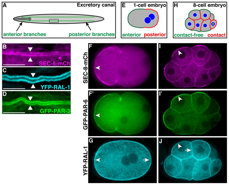
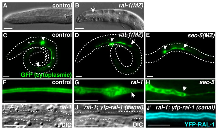
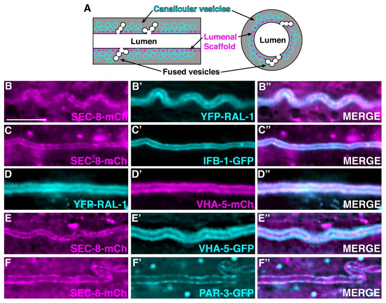
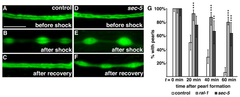
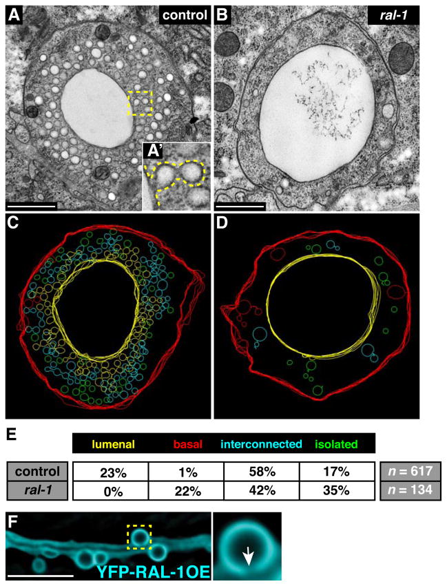
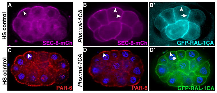
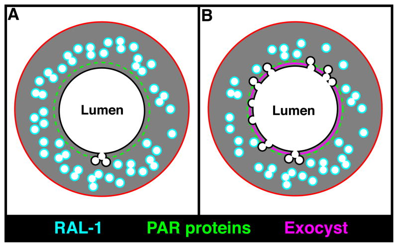
Lumenogenesis of small seamless tubes occurs through intracellular membrane growth and directed vesicle fusion events. Within the Caenorhabditis elegans excretory cell, which forms seamless intracellular tubes (canals) that mediate osmoregulation, lumens grow in length and diameter when vesicles fuse with the expanding lumenal surface. Here, we show that lumenal vesicle fusion depends on the small GTPase RAL-1, which localizes to vesicles and acts through the exocyst vesicle-tethering complex. Loss of either the exocyst or RAL-1 prevents excretory canal lumen extension. Within the excretory canal and other polarized cells, the exocyst co-localizes with the PAR polarity proteins PAR-3, PAR-6 and PKC-3. Using early embryonic cells to determine the functional relationships between the exocyst and PAR proteins, we show that RAL-1 recruits the exocyst to the membrane, while PAR proteins concentrate membrane-localized exocyst proteins to a polarized domain. These findings reveal that RAL-1 and the exocyst direct the polarized vesicle fusion events required for intracellular lumenogenesis of the excretory cell, suggesting mechanistic similarities in the formation of topologically distinct multicellular and intracellular lumens.

Keywords: Exocyst; Lumenogenesis; Osmoregulation; PAR proteins; Tubulogenesis; Vesicle trafficking.


Figures

Figure 1
Figure 1. RAL-1, exocyst and PAR protein expression in polarized cells
(A) Schematic of the excretory canal cell (green). The canal cell body and lateral branch are positioned adjacent the posterior pharynx (shaded dark gray). A representative region of posterior canal, depicted at higher magnification in B–D, is indicated by dashed rectangle. (B–D) Lateral view of excretory canal segment in L4 larvae expressing the indicated fusion proteins; arrowheads point towards canal lumen. (E) Schematic of a polarized 1-cell embryo displaying distinct anterior and posterior membrane domains. (F and F′) 1-cell embryo co-expressing SEC-8-mCherry (F) and PAR-6-GFP (F′), which are enriched at the anterior membrane (arrowheads). (G) 1-cell embryo expressing YFP-RAL-1, which localizes uniformly to both anterior (arrowhead) and posterior (arrow) membranes. (H) Schematic of a polarized 8-cell embryo with distinct contacted and contact-free cell surfaces; the germline precursor cell (asterisk) is unpolarized. (I and I′) 8-cell embryo co-expressing SEC-8-mCh (I) and PAR-6-GFP (I′), which are enriched at contact-free surfaces (arrowheads). (J) 8-cell embryo expressing YFP-RAL-1, which localizes uniformly to contact-free (arrowhead) and contacted (arrow) surfaces. In this figure and all subsequent figures, embryos and larvae are oriented anterior to the left, and whole embryos are ~50 μm in length. Scale bars in B–D are 10 μm.
Figure 2
Figure 2. ral-1 and the exocyst are required for osmoregulation and excretory canal lumen extension
(A–B) Anterior end of control (A) and ral-1(MZ) (B) L1 larvae placed in water; arrow indicates fluid-filled cavity. (C–E) Newly hatched L1 larvae (body outlined) expressing cytoplasmic GFP in the excretory canal cell. Arrowhead points to canal cell body, arrow indicates posterior extent of canal outgrowth, and asterisks in C highlight periodic cytoplasmic ‘pearls.’ (F–H) Segment of posterior excretory canal in L4 larvae of indicated genotypes expressing cytoplasmic GFP; mutant canals in G and H terminate prematurely (arrow). (I–J′) ral-1 mutant siblings lacking (I) or expressing (J and J′) canal-specific Ppgp-12::yfp-ral-1 transgene. Dashed lines in I and J outline the canal, which is discontinuous in ral-1 mutant (I) and smooth in rescued (J) canals; transgene expression in the rescued canal is shown in J′. Scale bars are 10 μm.
Figure 3
Figure 3. Subcellular localization of RAL-1, the exocyst, and PAR proteins within the excretory canal
(A) Excretory canal schematic shown in lateral (left) and transverse (right) sections. (B–F″) Live images of excretory canals in L4 larvae co-expressing the indicated transgenes. Scale bar is 10 μm, and scale is equivalent in all images.
Figure 4
Figure 4. ral-1 and sec-5 are required for prompt recovery from hyperosmotic shock
(A–C) Control larvae expressing canal-specific GFP before shock (A), 20 minutes after shock (B) and after one-hour recovery (C). (D–F) sec-5 mutant expressing canal-specific GFP; pearls remain after the one-hour recovery. (G) Percentage of control (n = 84), ral-1 (n = 55) and sec-5 (n = 33) mutants with cytoplasmic pearls following recovery from hyperosmotic shock; time zero corresponds to the first appearance of pearls following shock. Error bars represent the 95% confidence interval. **p < 0.001, *** p < 0.0001, Chi-squared test of mutants versus control. Scale bars are 10 μm, and scale is equivalent in all panels.
Figure 5
Figure 5. ral-1 is needed for vesicle fusion with the canal lumen
(A and A′) Representative transverse transmission electron microscopy (TEM) thin sections of the excretory canal in wild type (A) and ral-1 mutants (B); sections were taken from the posterior canal between the pharynx and distal gonad. A′ displays a higher magnification view of the boxed region in A; interconnected vesicles fused to the lumen are outlined by a dotted line. Scale bars in A and B are 500 nm. (C and D) Tracings from 200 nm thick section TEM tomograms (see Videos S1 and S2 for raw data Z-stacks) from regions immediately distal to the sections displayed in panels A and B. Each vesicle was outlined at its maximum diameter. Vesicles are classified as connected to the lumen (yellow), basal surface (red), interconnected (cyan), or isolated (green). (E) Summary of vesicle tracings from control (4 sections) and mutant (4 sections) tomograms; n refers to total number of vesicles from all sections combined. (F) L4 larval worm over-expressing YFP-RAL-1 specifically in the excretory canal. The magnified boxed region shows a cyst connected to the canal lumen (arrow). Scale bar is 10 μm.
Figure 6
Figure 6. PAR proteins in canal morphogenesis and exocyst polarity
(A–B′) Wild-type (A) and pkc-3ts mutant (B–B′) excretory canals from L4 larvae expressing GFP in the excretory canal and raised at 25°C after late embryogenesis; DIC image in B′ shows discontinuities of canal in B (compare to wild type in Figure S1D and to similar ral-1 mutant in Figure 2I). Scale bars are 10 μm. (C–D) 8-cell embryos from hermaphrodites expressing SEC-8-mCherry and fed with control bacteria containing empty vector (C) (n=66/66 polarized), or bacteria expressing pkc-3 dsRNA (n=19/20 not polarized). (E) pac-1 mutant 8-cell embryo expressing SEC-8-mCherry (n=16/16 not polarized). Arrowheads, SEC-8-mCherry at contact-free surfaces; arrows, SEC-8-mCherry at contacted surfaces. (F–G′) 26- to 28-cell stage control (n=24/24 polarized) (F) and mutant (n=13/13 polarized) (G, G′) embryos immunostained for indicated proteins. Dotted region in G and G′ outlines mutant cells depleted of maternally provided SEC-5-ZF1-YFP protein. Arrowheads, PAR-3 enriched at contact-free surfaces.
Figure 7
Figure 7. Effect of RAL-1CA on the exocyst and PAR proteins
(A–B′) Heat-shock (HS) control embryo (A) or sibling embryos expressing heat-shock driven GFP-RAL-1CA (B and B′); all embryos express SEC-8-mCherry, which is enriched at contact-free surfaces (arrowheads) in control embryos (n = 50/50 polarized) and recruited additionally to contacted surfaces (arrows) in embryos expressing RAL-1CA (n = 24/26 ectopically recruited). (C–D′) HS control embryo immunostained for PAR-6 (n=11/11 polarized) (C), and sibling embryo expressing heat-shock driven GFP-RAL-1CA (n=12/12 polarized) (D and D′) co-immunostained for PAR-6 and GFP-RAL-1CA. Arrowheads show PAR-6 enrichment at contact-free surfaces, arrow indicates contacted surfaces.
Figure 8
Figure 8. Model for excretory canal lumen formation
(A) Schematic of the excretory canal shown in transverse section, with the PAR proteins (green) and RAL-1 (cyan) present at distinct but overlapping domains. (B) At regions containing both PAR proteins and active RAL-1, the exocyst (magenta) is recruited and triggers the docking and fusion of canalicular vesicles with the lumenal surface, leading to canal expansion and increased exchange of water and salt.

References

    1. Achilleos A, Wehman AM, Nance J. PAR-3 mediates the initial clustering and apical localization of junction and polarity proteins during C. elegans intestinal epithelial cell polarization. Development. 2010;137:1833–1842. doi: 10.1242/dev.047647. - DOI - PMC - PubMed
    1. Anderson DC, Gill JS, Cinalli RM, Nance J. Polarization of the C. elegans embryo by RhoGAP-mediated exclusion of PAR-6 from cell contacts. Science. 2008;320:1771–1774. doi: 10.1126/science.1156063. - DOI - PMC - PubMed
    1. Andrew DJ, Ewald AJ. Morphogenesis of epithelial tubes: Insights into tube formation, elongation, and elaboration. Dev Biol. 2010;341:34–55. doi: 10.1016/j.ydbio.2009.09.024. - DOI - PMC - PubMed
    1. Boyd C. Vesicles carry most exocyst subunits to exocytic sites marked by the remaining two subunits, Sec3p and Exo70p. J Cell Biol. 2004;167:889–901. doi: 10.1083/jcb.200408124. - DOI - PMC - PubMed
    1. Bryant DM, Datta A, Rodríguez-Fraticelli AE, Peränen J, Martín-Belmonte F, Mostov KE. A molecular network for de novo generation of the apical surface and lumen. Nat Cell Biol. 2010;12:1035–1045. doi: 10.1038/ncb2106. - DOI - PMC - PubMed

Publication types

MeSH terms

LinkOut - more resources