Review
. 2015 Jan:36:49-71.
doi: 10.1016/j.yfrne.2014.07.001. Epub 2014 Aug 4.

Species, sex and individual differences in the vasotocin/vasopressin system: relationship to neurochemical signaling in the social behavior neural network

Affiliations
Review

Species, sex and individual differences in the vasotocin/vasopressin system: relationship to neurochemical signaling in the social behavior neural network

H Elliott Albers. Front Neuroendocrinol. 2015 Jan.

Abstract

Arginine-vasotocin (AVT)/arginine vasopressin (AVP) are members of the AVP/oxytocin (OT) superfamily of peptides that are involved in the regulation of social behavior, social cognition and emotion. Comparative studies have revealed that AVT/AVP and their receptors are found throughout the "social behavior neural network (SBNN)" and display the properties expected from a signaling system that controls social behavior (i.e., species, sex and individual differences and modulation by gonadal hormones and social factors). Neurochemical signaling within the SBNN likely involves a complex combination of synaptic mechanisms that co-release multiple chemical signals (e.g., classical neurotransmitters and AVT/AVP as well as other peptides) and non-synaptic mechanisms (i.e., volume transmission). Crosstalk between AVP/OT peptides and receptors within the SBNN is likely. A better understanding of the functional properties of neurochemical signaling in the SBNN will allow for a more refined examination of the relationships between this peptide system and species, sex and individual differences in sociality.

Keywords: Affiliation; Aggression; Communication; Estradiol; Oxytocin; Pair bonding; Sociality; Testosterone; V1a; V1b.


Figures

Figure 1
Figure 1
Confocal images of (A) medial paraventricular nucleus (mPVN), (B) medial supraoptic nucleus (mSON), and (C) nucleus circularis (NC) in a male Syrian hamster one hour after an agonistic encounter with another male. Green staining indicates arginine-vasopressin (AVP) and red staining indicates c-Fos. The agonistic encounter significantly increased AVP and c-Fos colocalization in the mSON and NC (p < 0.05) but not the mPVN (p > 0.05) as compared to controls (data not shown). From Terranova and Albers, unpublished data.
Figure 2
Figure 2
Variability in the microsatellite length of the V1a receptor gene in monogamous and promiscuous species of Peromyscus and Microtus. Top: Structure of the V1a gene: Green boxes represent microsatellites (MSAT 1 and MSAT 2) and brown regions represent Exons 1 and 2 (microsatellite 3 is not shown). Bottom: Allele sizes (basepairs) in eight species of Peromyscus (circles) and two species of Microtus (diamonds). Size of the homologous repeats were inferred from alignment of primers to GenBanK sequences (AF069304, AF070010) for comparison. Monogogamous species appear in blue and promiscuous appear in blue. Modified from (Turner and others, 2010).
Figure 3
Figure 3
Illustration of different patterns of synaptic release of ‘classical neurotransmitter’ (NT) and peptides. A: Moderate neuronal firing produces moderate levels of Ca2+ influx through voltage gated ion channels resulting in exocytosis of small synaptic vesicles (SSV; in purple) resulting in release of NT. B: High levels of neuronal firing produces high levels of Ca2+ influx through voltage gated ion channels resulting in exocytosis of both SSVs and large densecore vesicles (LDCV; in gray) resulting in release of NT and peptide. C: Two different peptides are packaged in LDCVs (red and blue) in a ratio of 1:1. High levels of neuronal firing produces high levels of Ca2+ influx through voltage gated ion channels resulting in exocytosis of SSVs and resulting in release of NT and exocytosis of LDCVs resulting the release of “cocktail” of peptides in a 1:1 ratio. D. Two different peptides are packaged in LDCVs (red and blue) in a ratio of 3:1. High levels of neuronal firing produces high levels of Ca2+ influx through voltage gated ion channels resulting in exocytosis of SSVs and resulting in release of NT and exocytosis of LDCVs resulting the release of “cocktail” of peptides in a 3:1 ratio.
Figure 4
Figure 4
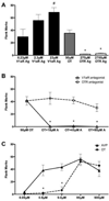
The induction of flank marking by intracerebroventricular injection of oxytocin (OT) is mediated by the vasopressin V1a receptor (V1aR). A. The amount of flank marking induced by the injection of the three concentrations of the selective V1aR agonist (Ag), [Phe2]OVT, OT or two concentrations of the selective OTR Ag, [Thr4,Gly7]OT, in 1 microliter of vehicle. (* and # indicate significant differences compared to the OT group) B. The amount of flank marking induced by the injection of 90 µM OT combined with vehicle or three concentrations of the selective V1a receptor antagonist (A), d(CH2)5[Tyr(Me)2]AVP (Manning Compound), or three concentrations of the selective OTR A, desGly-NH2-d(CH2)5[D-Tyr2,Thr4]OVT. (* indicates significant difference from the 90µM OT group). C. The amount of flank marking induced by the injection of various concentrations of OT and arginine-vasopressin (AVP) (* indicates significant difference between OT and AVP). From Song, McCann, McNeill, Larkin, Huhman and Albers, unpublished data.
Figure 5
Figure 5
Local network underlying the opposite effects of oxytocin (OT) and arginine-vasopressin (AVP) on fear. In the central amygdala (Ce) there are two adjacent but not overlapping populations of OT receptors (in the Ce lateral: CeL) and V1a receptors (in the Ce medial: CeM). In the CeL OT excites GABA containing inhibitory interneurons that synapse on neurons in the CeM that are excited by AVP and project to the brainstem. As a result, the effects of OT and AVP on fear are strikingly opposite. Modified from (Viviani and Stoop, 2008)
Figure 6
Figure 6
Hypothetical examples of how different combinations of synaptic and non-synaptic release of AVP-like peptides might regulate the expression of different social behaviors (i.e., X, Y or Z) by their action in the social behavior neural network. Blue lines represent neuronal activity in projections resulting in local synaptic release and green regions represent peptide released by volume transmission. See text for abbreviations.

References

    1. Absil P, Papello M, Viglietti-Panzica C, Balthazart J, Panzica G. The medial preoptic nucleus receives vasotocinergic inputs in male quail: a tract-tracing and immunocytochemical study. J Chem Neuroanat. 2002;24(1):27–39. - PubMed
    1. Acharjee S, Do-Rego JL, Oh DY, Moon JS, Ahn RS, Lee K, Bai DG, Vaudry H, Kwon HB, Seong JY. Molecular cloning, pharmacological characterization, and histochemical distribution of frog vasotocin and mesotocin receptors. J Mol Endocrinol. 2004;33(1):293–313. - PubMed
    1. Acher R, Chauvet J. The neurohypophyseal endocrine regulatory cascade: Precursors, mediators, receptors, and effectors. Front Neuroendocrinol. 1995;16:237–289. - PubMed
    1. Adkins-Regan E. Neuroendocrinology of social behavior. ILAR J. 2009;50(1):5–14. - PubMed
    1. Agnati LF, Fuxe K, Zoli M, Ozini I, Toffano G, Ferraguti F. A correlation analysis of the regional distribution of central enkephalin and beta-endorphin immunoreactive terminals and of opiate receptors in adult and old male rats. Evidence for the existence of two main types of communication in the central nervous system: the volume transmission and the wiring transmission. Acta Physiol Scand. 1986;128(2):201–207. - PubMed

Publication types