Review
. 2014 Oct:28:14-22.
doi: 10.1016/j.sbi.2014.07.003. Epub 2014 Aug 5.

Convergent and divergent mechanisms of sugar recognition across kingdoms

Affiliations
Review

Convergent and divergent mechanisms of sugar recognition across kingdoms

Maureen E Taylor et al. Curr Opin Struct Biol. 2014 Oct.

Abstract

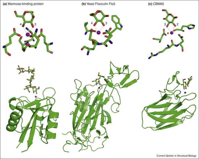
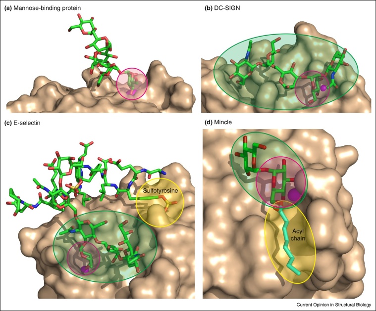
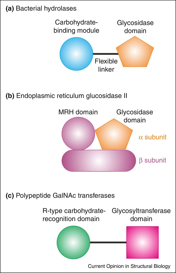
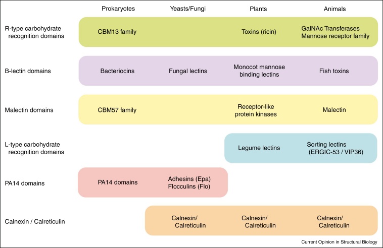
Protein modules that bind specific oligosaccharides are found across all kingdoms of life from single-celled organisms to man. Different, overlapping and evolving designations for sugar-binding domains in proteins can sometimes obscure common features that often reflect convergent solutions to the problem of distinguishing sugars with closely similar structures and binding them with sufficient affinity to achieve biologically meaningful results. Structural and functional analysis has revealed striking parallels between protein domains with widely different structures and evolutionary histories that employ common solutions to the sugar recognition problem. Recent studies also demonstrate that domains descended from common ancestors through divergent evolution appear more widely across the kingdoms of life than had previously been recognized.


Figures

Figure 1
Figure 1
Involvement of Ca2+ in sugar-binding sites in the context of multiple different protein folds. Top panels show close-up views of sugar-binding sites and lower panels show overall folds of carbohydrate-recognition domains. (a) Human serum mannose-binding protein (2MSB) with Man5 oligosaccharide. (b) Yeast flocculin Flo5 (2XJS) with bound mannobiose. (c) Family 60 carbohydrate-binding module (CBM60) from Cellvibrio japonicus xylanase (2XFD) with bound cellobiose. Ca2+ is indicated as a magenta sphere in each panel and water is represented as a red sphere. Coordination bonds from adjacent equatorial hydroxyl groups to the Ca2+ are indicated as dashed lines.
Figure 2
Figure 2
Multiple different ways in which binding specificity of C-type carbohydrate-recognition domains is enhanced by extended and accessory binding sites. Each of the binding sites involves a primary interaction between the Ca2+, shown in magenta, and two adjacent hydroxyl groups on a monosaccharide residue. (a) The relatively open binding site in mannose-binding protein binds only a terminal mannose residue, so only this residue interacts with the protein (2MSB). (b) DC-SIGN binds a more complex Man3GlcNAc2 oligosaccharide through an extended binding site that accommodates sugars on either side of the mannose residue in the primary binding site (1K9J). (c) In addition to ligation of fucose to Ca2+, the sialyl Lewisx oligosaccharide interacts with an extended binding site in E-selectin, which also has an accessory binding site for sulfated tyrosine residues on a glycoprotein ligand (1G1S). (d) Mincle binds to the disaccharide trehalose as a result of one glucose residue binding in the Ca2+ site and the second glucose residue contacting an adjacent site. In addition, glycolipid binding is enhanced through an accessory site that forms a hydrophobic grove which can interact with acyl chains on the 6-OH groups of the glucose residues (4KZV). Primary binding sites are highlighted in pink, extended oligosaccharide-binding sites are indicated in green and accessory sites for other modifications are shaded yellow.
Figure 3
Figure 3
Association of carbohydrate-recognition domains with enzymatically active domains. (a) One or more carbohydrate-binding modules are often linked to bacterial glycosidases and cellulose-degrading enzymes in a single polypeptide. The carbohydrate-binding modules localize the activity on substrates and enhance the activity of enzymes. (b) The α subunit of endoplasmic reticulum glucosidase II contains the glucosidase active site, but the activity of the enzyme on high mannose oligosaccharides that bear terminal glucose residues on one branch is enhanced by the β subunit, which contains an MRH domain that binds mannose on another branch of the oligosaccharide. (c) R-type carbohydrate-recognition domains in many of the polypeptide GalNAc transferase proteins direct the enzyme to regions of substrate glycoproteins that already bear one or more GalNAc residues.
Figure 4
Figure 4
A summary of some of the types of carbohydrate-recognition domains that are found in a wide range of species. Other types of domain not shown are expressed only in more restricted groups of organisms. For example, galectins, siglecs and C-type lectins are expressed only in animals and several classes of adhesins are specific to bacteria.

References

    1. Sharon N., Lis H. History of lectins: from hemagglutinins to biological recognition molecules. Glycobiology. 2004;14:53R–62R. - PubMed
    1. Etzold S., Juge N. Structural insights into bacterial recognition of intestinal mucins. Curr Opin Struct Biol. 2014;28 (in press) - PubMed
    1. Abbott W.D., van Bueren A.L. Using structure to inform carbohydrate binding module function. Curr Opin Struct Biol. 2014;28 (in press) - PubMed
    1. Drickamer K., Taylor M.E. Identification of lectins from genomic sequence data. Methods Enzymol. 2003;362:592–599. - PubMed
    1. Weis W.I., Taylor M.E., Drickamer K. The C-type lectin superfamily in the immune system. Immunol Rev. 1998;163:19–34. - PubMed

Publication types