. 2014 Oct 1;25(19):2984-92.
doi: 10.1091/mbc.E13-09-0514. Epub 2014 Aug 7.

The Caenorhabditis elegans pericentriolar material components SPD-2 and SPD-5 are monomeric in the cytoplasm before incorporation into the PCM matrix

Affiliations

The Caenorhabditis elegans pericentriolar material components SPD-2 and SPD-5 are monomeric in the cytoplasm before incorporation into the PCM matrix

Oliver Wueseke et al. Mol Biol Cell. .

Abstract

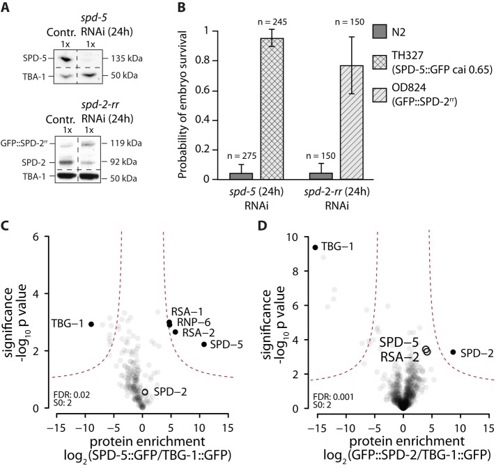
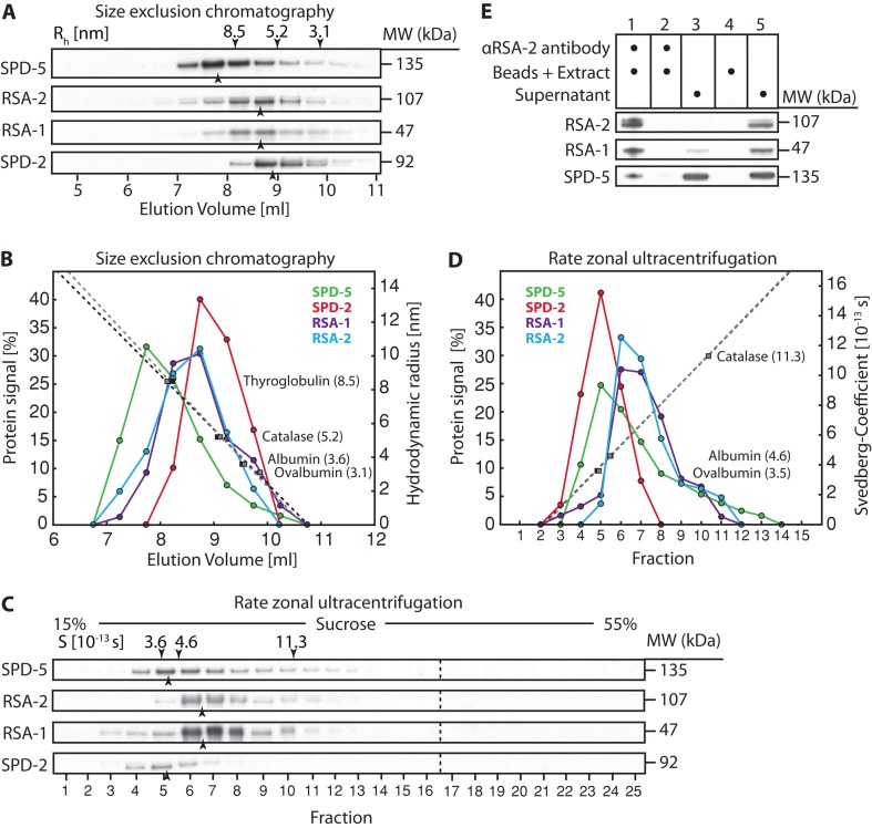
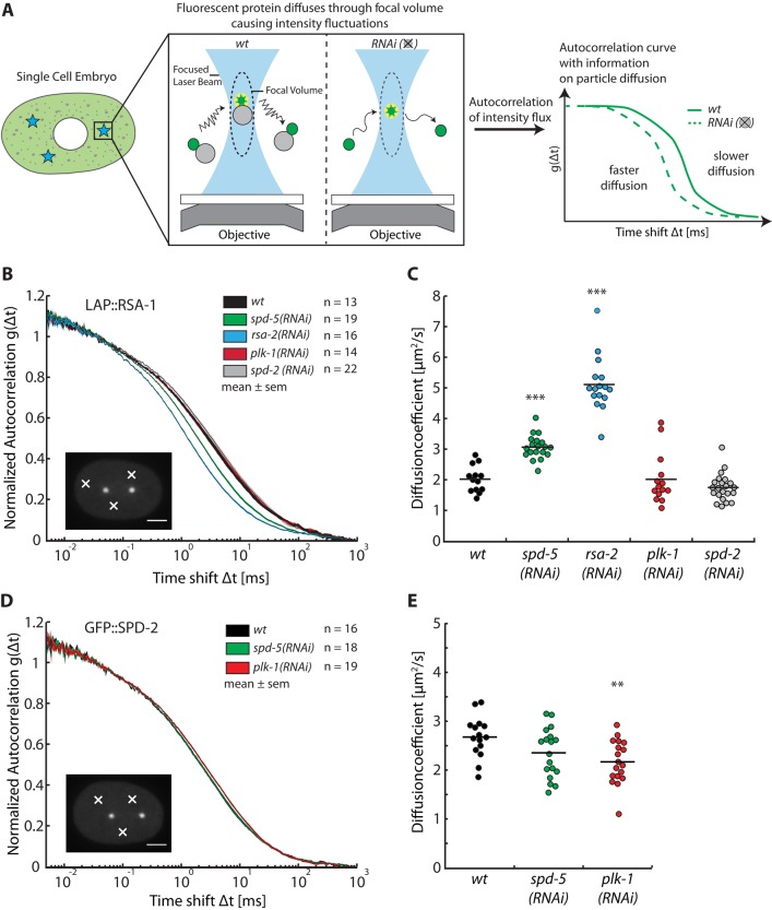
Centrosomes are the main microtubule-organizing centers in animal cells. Centrosomes consist of a pair of centrioles surrounded by a matrix of pericentriolar material (PCM) that assembles from cytoplasmic components. In Caenorhabditis elegans embryos, interactions between the coiled-coil proteins SPD-5 and SPD-2 and the kinase PLK-1 are critical for PCM assembly. However, it is not known whether these interactions promote the formation of cytoplasmic complexes that are added to the PCM or whether the components interact only during incorporation into the PCM matrix. Here we address this problem by using a combination of live-cell fluorescence correlation spectroscopy, mass spectrometry, and hydrodynamic techniques to investigate the native state of PCM components in the cytoplasm. We show that SPD-2 is monomeric, and neither SPD-2 nor SPD-5 exists in complex with PLK-1. SPD-5 exists mostly as a monomer but also forms complexes with the PP2A-regulatory proteins RSA-1 and RSA-2, which are required for microtubule organization at centrosomes. These results suggest that the interactions between SPD-2, SPD-5, and PLK-1 do not result in formation of cytoplasmic complexes, but instead occur in the context of PCM assembly.


Figures

FIGURE 1:
FIGURE 1:
Identification of cytoplasmic interactors of GFP::SPD-2 and SPD-5::GFP using label-free quantification mass spectrometry. (A) Western blot showing depletion of endogenous SPD-5 or SPD-2 after 24 h of RNAi treatment. SPD-5 is fully removed from N2 worms after 24 h of RNAi. Endogenous SPD-2 levels in OD824 (GFP::SPD-2-rr) are reduced to 25% after 24 h of spd-2-rr RNAi, whereas GFP::SPD-2-rr levels increase threefold. (B) Embryonic viability of N2, OD824 (GFP::SPD-2rr), and TH327 (SPD-5::GFP codon optimized to cai 0.65) embryos after 24 h of treatment with spd-5 or spd-2-rr RNAi. Both transgenes were found to restore embryo viability when endogenous protein was removed. (C, D) Identification of cytoplasmic interactors of SPD-5::GFP and GFP::SPD-2. SPD-5::GFP or GFP::SPD-2 was immunopurified from C. elegans embryo extracts, and the coimmunoprecipitated proteins were identified via label-free quantification mass spectrometry. TBG-1::GFP immunoprecipitates were used as a control. Experiments were repeated in triplicate. Results are plotted as volcano plots, with the enrichment of identified peptides in sample vs. control plotted on the x-axis and the significance of enrichment on the y-axis. p values and enrichments were calculated as described previously (Hubner et al., 2010). Black circles indicate nonenriched centrosome proteins; black dots indicate the control protein, as well as significant interactors enriched in the sample.
FIGURE 2:
FIGURE 2:
Stoichiometry and shape analysis of SPD-2, SPD-5, and the SPD-5/RSA-1/RSA-2 complex. (A) Cytoplasmic extracts prepared from C. elegans embryos were fractionated by size exclusion chromatography, and an immunoblot of the fractions (collected in 0.5-ml steps) probed for SPD-5, RSA-2, RSA-1, and SPD-2 is shown. Black arrowheads mark the center of the peak for each protein. The centers of the peaks for each standard protein are indicated, along with their hydrodynamic radius, by the black arrowheads above the top lane. (B) Graph blotting the measured signal for each protein vs. elution volume. ­Standard calibration curves were repeated twice (dotted lines) and calculated from the position of the standard proteins (filled rectangles). (C) Svedberg coefficients for SPD-5, RSA-1, RSA-2, and SPD-2 were determined by rate zonal ultracentrifugation. An immunoblot of the 25 × 0.2 ml fractions obtained from a 5-ml 15–55% sucrose gradient is shown. Black arrowheads mark the center of the peak for each protein. The centers of the peaks for each standard protein are indicated, along with their Svedberg coefficients, by the black arrowheads above the top lane. (D) Graph plotting the measured signal for each protein vs. fraction. The standard calibration curve (dotted line) was calculated from the position of the standard proteins (filled rectangles). (E) Immunoblot of RSA-2 coimmunoprecipitation experiment using RSA-2–specific antibodies on cytoplasmic extract, showing that SPD-5 and RSA-1 coimmunoprecipitate with RSA-2. 1) αRSA-2 antibody–coupled beads were incubated with fresh cytoplasmic extract; 2) the remaining extract from step 1 was reincubated with fresh αRSA-2 antibody–coupled beads to control for full depletion of RSA-2; 3) remaining supernatant from step 2; 4) blank beads were incubated with fresh cytoplasmic extract to control for unspecific binding of beads; 5) remaining supernatant from step 4.
FIGURE 3:
FIGURE 3:
Fluorescence correlation spectroscopy reveals cytoplasmic interactions in vivo. (A) Schematic overview of the experimental procedure. FCS measurements were conducted in the cytoplasm of single-cell embryos at three randomly chosen cytoplasmic positions away from the centrosomes and nucleus. Intensity fluctuations caused by GFP-tagged proteins diffusing through the focal volume were recorded over time. (B) Diffusion of LAP::RSA-1 is affected by depletion or RSA-2 and SPD-5. Autocorrelation curves are depicted as mean ± SEM as calculated from all embryos for each condition. The number of embryos analyzed is indicated next to each condition. Scale bar, 10 μm; white crosses point to exemplary locations of measurements. (C) Diffusion coefficient plot showing diffusion coefficients of LAP::RSA-1. LAP::RSA-1 diffusion changes from 2.0 ± 0.4 to 3.1 ± 0.4 μm2/s (p < 0.001, n = 19) under spd-5 RNAi and 5.1 ± 0.9 μm2/s (p < 0.001, n = 16) under rsa-2 RNAi. Diffusion coefficients were obtained from fitting the autocorrelation curve of LAP::RSA-1 from each embryo. Asterisks indicate significance with p < 0.001. The results show a clear interaction between LAP::RSA-1 and RSA-2, as well as SPD-5. An interaction with PLK-1 or SPD-2 could not be detected. (D) GFP::SPD-2 diffusion is unaffected by depletion of SPD-5 but slightly decreases under PLK-1 depletion. Representation as in B, showing GFP::SPD-2 autocorrelation curves under various conditions. (E) Diffusion coefficient plot of GFP::SPD-2. GFP::SPD-2 diffuses indistinguishably, with ∼2.5 μm2/s in wild- type and spd-5 RNAi conditions. Its diffusion is significantly reduced to ∼2.2 μm2/s under plk-1 RNAi conditions.

References

    1. Bacia K, Kim SA, Schwille P. Fluorescence cross-correlation spectroscopy in living cells. Nat Methods. 2006;3:83–89. - PubMed
    1. Bacia K, Schwille P. Practical guidelines for dual-color fluorescence cross-correlation spectroscopy. Nat Protoc. 2007;2:2842–2856. - PubMed
    1. Banks DS, Fradin C. Anomalous diffusion of proteins due to molecular crowding. Biophys J. 2005;89:2960–2971. - PMC - PubMed
    1. Baudendistel N, Müller G, Waldeck W. 2005. Two-hybrid fluorescence cross-correlation spectroscopy detects protein–protein interactions in vivo Chemphyschem 6, 984–990. - PubMed
    1. Boxem M, Maliga Z, Klitgord N, Li N, Lemmens I, Mana M, de Lichtervelde L, Mul J, van de Peut D, Devos M. A protein domain-based interactome network for C. elegans early embryogenesis. Cell. 2008;134:534–545. - PMC - PubMed

Publication types

MeSH terms