. 2014 Aug 7;55(9):5497-509.
doi: 10.1167/iovs.14-14519.

Differential effects of caveolin-1 and -2 knockdown on aqueous outflow and altered extracellular matrix turnover in caveolin-silenced trabecular meshwork cells

Affiliations

Differential effects of caveolin-1 and -2 knockdown on aqueous outflow and altered extracellular matrix turnover in caveolin-silenced trabecular meshwork cells

Mini Aga et al. Invest Ophthalmol Vis Sci. .

Abstract

Purpose: A single nucleotide polymorphism (SNP) identified between caveolin-1 (CAV1) and caveolin-2 (CAV2) on chromosome 7 is associated with glaucoma. One function of CAVs is endocytosis and recycling of extracellular matrix (ECM) components. Here, we generated CAV-silencing lentivirus to evaluate the effects on ECM turnover by trabecular meshwork (TM) cells and to measure the effect on outflow facility in anterior segment perfusion culture.

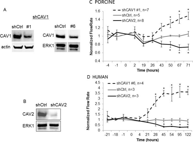
Methods: Short hairpin CAV1 and CAV2 silencing and control lentivirus were generated, characterized, and applied to anterior segments in perfusion culture. Colocalization of CAVs with various ECM molecules in TM cells was investigated using immunofluorescence and confocal microscopy. Western immunoblotting and fluorogenic-based enzyme activity assays were used to investigate ECM protein levels and degradation, respectively.

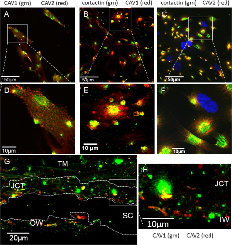
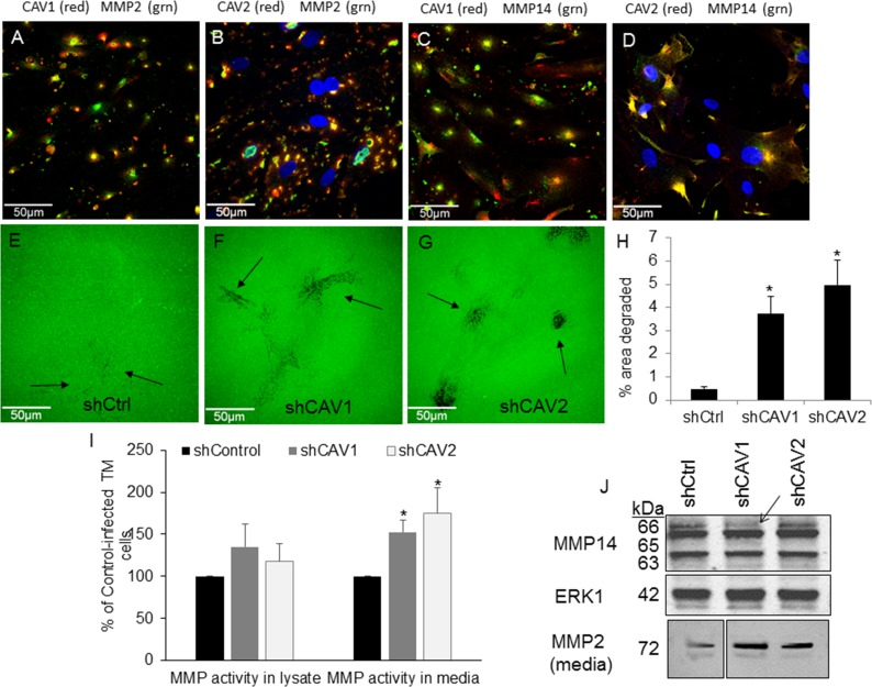
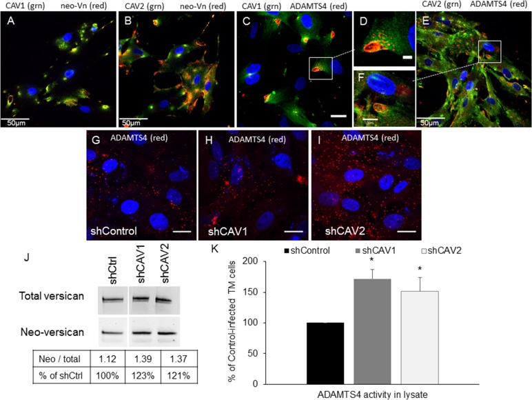
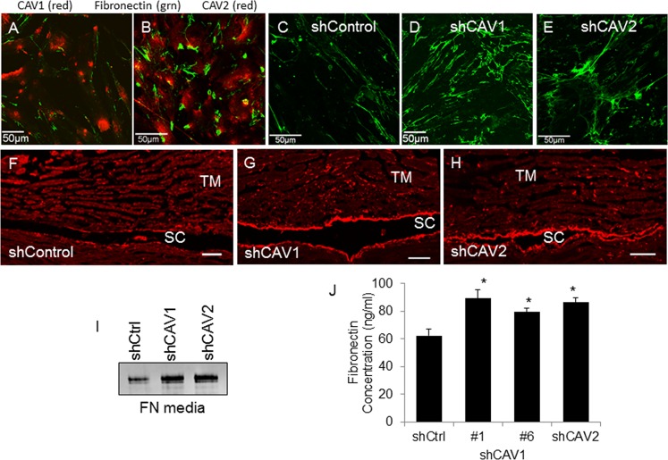
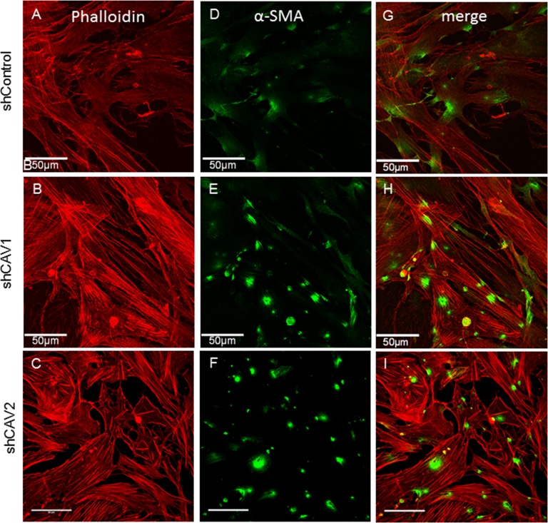
Results: Endogenous CAVs colocalized with cortactin at podosome- or invadopodia-like structures (PILS), which are areas of focal ECM degradation. In perfusion culture, outflow rates increased significantly in CAV1-silenced anterior segments, whereas outflow significantly decreased in CAV2-silenced anterior segments. Matrix metalloproteinase (MMP)2 and MMP14, and a disintegrin and metalloproteinase with thrombospondin motifs-4 (ADAMTS4) colocalized with both CAVs in TM cells. Protein levels and enzyme activities of MMP/ADAMTS4, fibronectin protein levels, actin stress fibers, and α-smooth muscle actin were all increased in CAV-silenced cells.

Conclusions: Caveolin-mediated endocytosis is one mechanism by which TM cells can alter the physiological catabolism of ECM in order to change the composition of the outflow channels in the TM to regulate aqueous outflow resistance. Dysregulation of CAV function could contribute to the pathological changes in ECM that are observed in glaucoma.

Keywords: endocytosis; extracellular matrix; glaucoma anterior segment; matrix metalloproteinases; trabecular meshwork.


Figures

Figure 1
Figure 1
Colocalization of caveolins-1 and -2 with each other and with cortactin, a biomarker of PILS. (A) Localization of CAV1 (green) and CAV2 (red) in human TM cells. (B) Caveolin-1 (red) localized with cortactin (green). (C) Caveolin-2 (red) localized with cortactin (green). We used DAPI to stain nuclei blue. (DF) Show higher magnification images of the boxed areas in (AC). (G) A frontal section of human TM tissue immunostained with CAV1 (green) and CAV2 (red). Partial colocalization was observed, particularly in the JCT region adjacent to Schlemm's canal. The TM beams show slight green autofluorescence. A dotted line delineates the wall of SC and another dotted line shows approximately the depth of the JCT region (7–20 μm from the IW). (H) Shows a higher magnification image of the boxed area in (G). Scale bars: (AC) 50 μm; (G) 20 μm; (DF, H) 10 μm.
Figure 2
Figure 2
Caveolin shRNA gene silencing. (A) Western immunoblotting shows a significant decrease in CAV1 protein in shCAV1-infected compared with control (shCtrl) in TM cells. Actin or total ERK1 loading controls are shown. (B) Western immunoblotting shows a significant decrease in CAV2 protein in control (shCtrl) and shCAV2-infected TM cells. Total ERK1 loading control is shown. In perfused porcine (C) and human (D) anterior segments, shCAV1 significantly increased outflow rates over time, while shCAV2 decreased outflow rates. Time 0 indicates the time of application of lentivirus. Data show the average flow rate ± SEM; n is indicated in the graph legend. *P < 0.05 by ANOVA.
Figure 3
Figure 3
Effects of CAV silencing on MMP levels and activity. Localization of endogenous CAV1 ([A, C], red) or CAV2 ([B, D]; red) compared with MMP2 ([A, B]; green) or MMP14 ([C, D]; green) are shown in human TM cells. We used DAPI to stain nuclei blue. (EG) Degradation of Oregon Green 488–conjugated gelatin (Life Technologies) 2 hours after plating control (E), CAV1- (F), and CAV2-silenced (G) human TM cells. Arrows point to areas of degradation. Scale bars: 50 μm. (H) Quantitative analysis of the areas of gelatin degradation using Fiji image processing software. Data is averaged (±SEM) from multiple images: n = 5 for shControl; n = 9 for shCAV1; n = 6 for shCAV2. *P < 0.01. (I) Activity of MMP was measured in cell lysates and media using a fluorogenic MMP assay with a generic peptide substrate. Results show RFUs normalized to total protein in each sample and calculated as a percentage of control. Error bars represent SEM. *P < 0.05 by ANOVA; n = 5. (J) Western immunoblots of MMP14 in cell lysates and MMP2 in media of control- and CAV-infected TM cells. Arrow points to a reduction of the proform of MMP14 in shCAV1-silenced TM cells. Total ERK1 was used as a loading control for lysates. For the media lanes, equal amounts of total protein were loaded. Positions of molecular weight markers are shown in kDa.
Figure 4
Figure 4
Effects of CAV silencing on ADAMTS4 immunolocalization and activity. Colocalization of endogenous CAV1 ([A, C, D]; green) and CAV2 ([B, E, F]; green) with versican neoepitope ([A, B]; red) and ADAMTS4 ([CF]; red). Versican neoepitope (neo-Vn) is exposed following proteolytic cleavage by ADAMTS4. (GI) Immunostaining with ADAMTS4 immunostaining of control (G), CAV1- (H) or CAV2-silenced (I) human TM cells. We used DAPI to stain nuclei blue. Scale bars: (A, B, E) 50 μm; (C, GI) 20 μm; (F) 10 μm; (D) 5 μm. (J) Western immunoblots showing total versican and neo-Vn epitope bands in control and CAV-silenced TM cells. The Table shows the densitometry ratio of neoepitope to total versican for each condition and then calculated as a percentage of the control. (K) ADAMTS4 activity in cell lysates was quantified by a fluorogenic enzyme activity assay. Results show RFUs normalized to total protein in each sample. Error bars represent SEM. *P < 0.05 by ANOVA; n = 5.
Figure 5
Figure 5
Effects of CAV silencing on fibronectin. Colocalization of endogenous CAV1 ([A]; red) and CAV2 ([B]; red) with fibronectin (green). Fibronectin immunostaining in control (C, F), CAV1- (D, G) and CAV2-silenced (E, H) TM cells (CE) and radial sections of perfused human anterior segments (FH). Confocal acquisition settings were identical for control and CAV-silenced cells. Scale bars: (AE) 50 μm; (FH) 20 μm. (I) Western immunoblot analysis of fibronectin levels in the media of control and CAV-silenced TM cells. Equal amounts of protein were loaded into each lane. (J) A fibronectin ELISA assay of control and CAV-silenced human TM cell lysates. Data is the mean ± SEM; n = 3. *P < 0.05 compared with shControl by ANOVA.
Figure 6
Figure 6
Effects of CAV silencing on the actin cytoskeleton. Human TM cells were infected with control (A, D, G), CAV1-silencing (B, E, H), or CAV2-silencing (C, F, I) lentivirus. F-actin was stained with phalloidin-594 (red; [AC], [GI]) and αSMA immunostaining of CAV-silenced TM cells (green; [DF], [GI]). The merged images are shown (GI). Confocal acquisition settings were identical for control and CAV-silenced cells. Scale bars: 50 μm.

References

    1. Boland MV, Quigley HA. Risk factors and open-angle glaucoma: classification and application. J Glaucoma. 2007; 16: 406–418 - PubMed
    1. Rezaie T, Child A, Hitchings R, et al. Adult-onset primary open-angle glaucoma caused by mutations in optineurin. Science. 2002; 295: 1077–1079 - PubMed
    1. Stone EM, Fingert JH, Alward WL, et al. Identification of a gene that causes primary open angle glaucoma. Science. 1997; 275: 668–670 - PubMed
    1. Monemi S, Spaeth G, DaSilva A, et al. Identification of a novel adult-onset primary open-angle glaucoma (POAG) gene on 5q22.1. Hum Mol Genet. 2005; 14: 725–733 - PubMed
    1. Pasutto F, Keller KE, Weisschuh N, et al. Variants in ASB10 are associated with open-angle glaucoma. Hum Mol Genet. 2012; 21: 1336–1349 - PMC - PubMed

Publication types