

Single molecule investigation of Ag+ interactions with single cytosine-, methylcytosine- and hydroxymethylcytosine-cytosine mismatches in a nanopore
- PMID: 25103463
- PMCID: PMC4126007
- DOI: 10.1038/srep05883
Single molecule investigation of Ag+ interactions with single cytosine-, methylcytosine- and hydroxymethylcytosine-cytosine mismatches in a nanopore
Erratum in
-
Corrigendum: Single Molecule Investigation of Ag+ Interactions with Single Cytosine-, Methylcytosine- and Hydroxymethylcytosine-Cytosine Mismatches in a Nanopore.Sci Rep. 2015 Jul 10;5:9732. doi: 10.1038/srep09732. Sci Rep. 2015. PMID: 26161846 Free PMC article. No abstract available.
Abstract
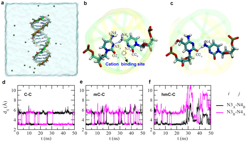
Both cytosine-Ag-cytosine interactions and cytosine modifications in a DNA duplex have attracted great interest for research. Cytosine (C) modifications such as methylcytosine (mC) and hydroxymethylcytosine (hmC) are associated with tumorigenesis. However, a method for directly discriminating C, mC and hmC bases without labeling, modification and amplification is still missing. Additionally, the nature of coordination of Ag(+) with cytosine-cytosine (C-C) mismatches is not clearly understood. Utilizing the alpha-hemolysin nanopore, we show that in the presence of Ag(+), duplex stability is most increased for the cytosine-cytosine (C-C) pair, followed by the cytosine-methylcytosine (C-mC) pair, and the cytosine-hydroxymethylcytosine (C-hmC) pair, which has no observable Ag(+) induced stabilization. Molecular dynamics simulations reveal that the hydrogen-bond-mediated paring of a C-C mismatch results in a binding site for Ag(+). Cytosine modifications (such as mC and hmC) disrupted the hydrogen bond, resulting in disruption of the Ag(+) binding site. Our experimental method provides a novel platform to study the metal ion-DNA interactions and could also serve as a direct detection method for nucleobase modifications.
Figures


Similar articles
-
Discrimination of methylcytosine from hydroxymethylcytosine in DNA molecules.J Am Chem Soc. 2011 Jan 26;133(3):486-92. doi: 10.1021/ja107836t. Epub 2010 Dec 14. J Am Chem Soc. 2011. PMID: 21155562 Free PMC article.
-
Identification of epigenetic DNA modifications with a protein nanopore.Chem Commun (Camb). 2010 Nov 21;46(43):8195-7. doi: 10.1039/c0cc02864a. Epub 2010 Oct 6. Chem Commun (Camb). 2010. PMID: 20927439 Free PMC article.
-
High sensitivity 5-hydroxymethylcytosine detection in Balb/C brain tissue.J Vis Exp. 2011 Feb 1;(48):2661. doi: 10.3791/2661. J Vis Exp. 2011. PMID: 21307836 Free PMC article.
-
New themes in the biological functions of 5-methylcytosine and 5-hydroxymethylcytosine.Immunol Rev. 2015 Jan;263(1):36-49. doi: 10.1111/imr.12242. Immunol Rev. 2015. PMID: 25510270 Free PMC article. Review.
-
High-throughput sequencing offers new insights into 5-hydroxymethylcytosine.Biomol Concepts. 2016 Jun 1;7(3):169-78. doi: 10.1515/bmc-2016-0011. Biomol Concepts. 2016. PMID: 27356236 Free PMC article. Review.
Cited by
-
Real-time label-free detection of dynamic aptamer-small molecule interactions using a nanopore nucleic acid conformational sensor.Proc Natl Acad Sci U S A. 2023 Jun 13;120(24):e2108118120. doi: 10.1073/pnas.2108118120. Epub 2023 Jun 5. Proc Natl Acad Sci U S A. 2023. PMID: 37276386 Free PMC article.
-
Nanopore sensing of botulinum toxin type B by discriminating an enzymatically cleaved Peptide from a synaptic protein synaptobrevin 2 derivative.ACS Appl Mater Interfaces. 2015 Jan 14;7(1):184-92. doi: 10.1021/am5056596. Epub 2014 Dec 29. ACS Appl Mater Interfaces. 2015. PMID: 25511125 Free PMC article.
-
Remote Activation of a Nanopore for High-Performance Genetic Detection Using a pH Taxis-Mimicking Mechanism.Anal Chem. 2017 Dec 19;89(24):13039-13043. doi: 10.1021/acs.analchem.7b03979. Epub 2017 Dec 4. Anal Chem. 2017. PMID: 29183111 Free PMC article.
-
Characterization of Interstrand DNA-DNA Cross-Links Using the α-Hemolysin Protein Nanopore.ACS Nano. 2015 Dec 22;9(12):11812-9. doi: 10.1021/acsnano.5b03923. Epub 2015 Nov 18. ACS Nano. 2015. PMID: 26563913 Free PMC article.
-
Dynamics of a DNA Mismatch Site Held in Confinement Discriminate Epigenetic Modifications of Cytosine.J Am Chem Soc. 2017 Feb 22;139(7):2750-2756. doi: 10.1021/jacs.6b12284. Epub 2017 Feb 13. J Am Chem Soc. 2017. PMID: 28125225 Free PMC article.
References
-
- Lin Y. H. & Tseng W. L. Highly sensitive and selective detection of silver ions and silver nanoparticles in aqueous solution using an oligonucleotide-based fluorogenic probe. Chem Commun 43, 6619–21 (2009). - PubMed
-
- Ono A. et al. Specific interactions between silver(I) ions and cytosine-cytosine pairs in DNA duplexes. Chem Commun 39, 4825–7 (2008). - PubMed
-
- Wen Y. et al. A graphene-based fluorescent nanoprobe for silver(I) ions detection by using graphene oxide and a silver-specific oligonucleotide. Chem Commun 46, 2596–8 (2010). - PubMed
-
- Torigoe H. et al. Thermodynamic and structural properties of the specific binding between Ag(+) ion and C:C mismatched base pair in duplex DNA to form C-Ag-C metal-mediated base pair. Biochimie 94, 2431–40 (2012). - PubMed
-
- Miyake Y. et al. MercuryII-mediated formation of thymine-HgII-thymine base pairs in DNA duplexes. J Am Chem Soc 128, 2172–3 (2006). - PubMed
Publication types
MeSH terms
Substances
Grants and funding
LinkOut - more resources
Full Text Sources
Other Literature Sources
Molecular Biology Databases
