. 2014 Aug 7:14:61.
doi: 10.1186/1471-2490-14-61.

Interpositional substitution of free vas deferens segment autografts in rat: feasibility and potential implications

Affiliations

Interpositional substitution of free vas deferens segment autografts in rat: feasibility and potential implications

Teoman Cem Kadioglu et al. BMC Urol. .

Abstract

Background: Insufficient vas length for performing a tension-free vasovasostomy is a problem occasionally encountered by microsurgeons. Herein we evaluated utilization of a non-vascularized vas deferens autograft in a rat model.

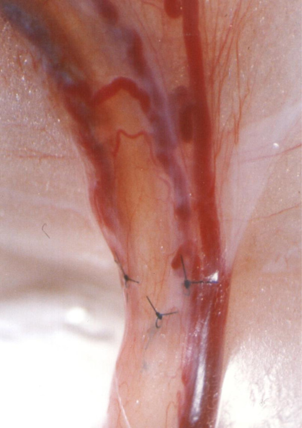
Methods: Segments of isolated vas deferens, 2.5 cm in length, were used as bilateral autografts in 15 rats. Each autograft was implanted between the two transected ends of vas deferens using end-to-end anastomosis. Fertility, sperm motility, and graft survival was evaluated and compared with the control group.

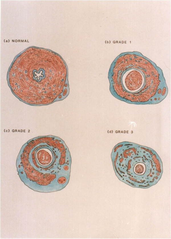
Results: At the end of the 3 months, 9/15 (60%) rats were able to breed successfully and 24 (80%) vas grafts were patent and viable. Large granulomata developed at the proximal anastomosis sites in 6 (20%) autografts that failed. Unilateral minimal fluid leakage was observed in 6 (20%) of the proximal (testicular end) anastomosis sites in those rats that were able to breed. Histological evaluations demonstrated that graft survival was associated with mild to severe changes in the structure of the vas autograft. On semen analysis 76% of the sperms in the experimental group had forward motility compared to 78% in the control group (p > 0.05).

Conclusions: Vas autograft can successfully be performed in a rat model with ultimate breeding capability.


Figures

Figure 1
Figure 1
Vas autograft microsurgical procedure in the rat. (a) Vas preparation prior to transaction showing a loop of exposed intact straight vas (v) with transaction sites marked (dashed lines). e, epididymis; f, epididymal fat pad; cv, convoluted vas. (b) Isolated 2.5 cm vas segment prepared for use as an autograft. Note the increasing diameter from proximal (p) to distal (d), which is associated with the increasing thickness of the smooth muscle layers. (c) Vas autograft (vg) in position after microsurgery showing the proximal (p) and distal (d) lines of 10/0 sutures along each anastomosis site.
Figure 2
Figure 2
Grading system. (a) Grade 0: Control vas graft; normal histology. (b) Grade 1: Loss of stellate lumen but maintenance of periluminal smooth muscle continuity. (c) Grade 2: Reduction in mass and progressive fibrosis of the longitudinal and circular smooth muscle causing discontinuity of muscle layers. (d) Grade 3: Smooth muscle layers are largely replaced by fibrous tissue surrounded by a revascularizing, thickened adventitia.
Figure 3
Figure 3
Longitudinal section through the proximal suture line of a vas autograft showing the patency of the anastomosis. The spermatozoa (s) filling the lumen, the suture (S), and the interrupted smooth muscle (sm) layers along the anastomosis site (dashed lines) are shown. Proximal vas (p), vas autograft (vg). Note the reduced length of the stereocilia (arrows) in the vas autograft 135x.
Figure 4
Figure 4
Minimal leakage at the proximal anastomosis site.
Figure 5
Figure 5
Prominent neovascularity.
Figure 6
Figure 6
Normal vas deferens.
Figure 7
Figure 7
Transverse section through a vas autograft 1 month after surgery showing minimal changes. The slightly dilated lumen (l) has partially lost its stellate shape and contains few spermatozoa, and the smooth muscle (sm) layers have remained intact with thickened, revascularized adventitia (arrows). Note the presence of stereocilia on the epithelial cells as in controls 55x.
Figure 8
Figure 8
Transverse section through a vas autograft 1 month after surgery showing mild degenerative changes. The enlarged, rounded lumen (l) exhibits an increased presence of spermatozoa (s), and disrupted smooth muscle (sm) layers are partially replaced by fibrous tissue (f) surrounded by a revascularizing, thickened adventitia (arrows). Note the reduced epithelial height (arrow heads) and large perivasal pad of adipose tissue (a) 55x.
Figure 9
Figure 9
Transverse section through a vas autograft 1 month after surgery showing severe degenerative changes. The rounded lumen (l) exhibits an increased presence of spermatozoa (s), and smooth muscle (arrow heads) layers are largely replaced by fibrous tissue (f) surrounded by a revascularizing, thickened adventitia (arrows) 55x.

References

    1. Koslov DS, Andersson KE. Physiological and pharmacological aspects of the vas deferens-an update. Front Pharmacol. 2013;4:101. - PMC - PubMed
    1. Rajfer J, Bennett CJ. Vasectomy. Urol Clin North Am. 1988;15:631. - PubMed
    1. Kelami A. A new concept for macroscopic anastomosis of vas deferens; suture material and operative technique. Andrologia. 1980;12(3):245. - PubMed
    1. Bernie AM, Osterberg EC, Stahl PJ, Ramasamy R, Goldstein M. Vasectomy reversal in humans. Spermatogenesis. 2012;2(4):273–278. doi: 10.4161/spmg.22591. - DOI - PMC - PubMed
    1. Parekattil SJ, Gudeloglu A. Robotic assisted andrological surgery. Asian J Androl. 2013;15(1):67–74. doi: 10.1038/aja.2012.131. - DOI - PMC - PubMed

LinkOut - more resources