

Melanocyte stem cells as potential therapeutics in skin disorders
- PMID: 25104310
- PMCID: PMC4616011
- DOI: 10.1517/14712598.2014.935331
Melanocyte stem cells as potential therapeutics in skin disorders
Abstract
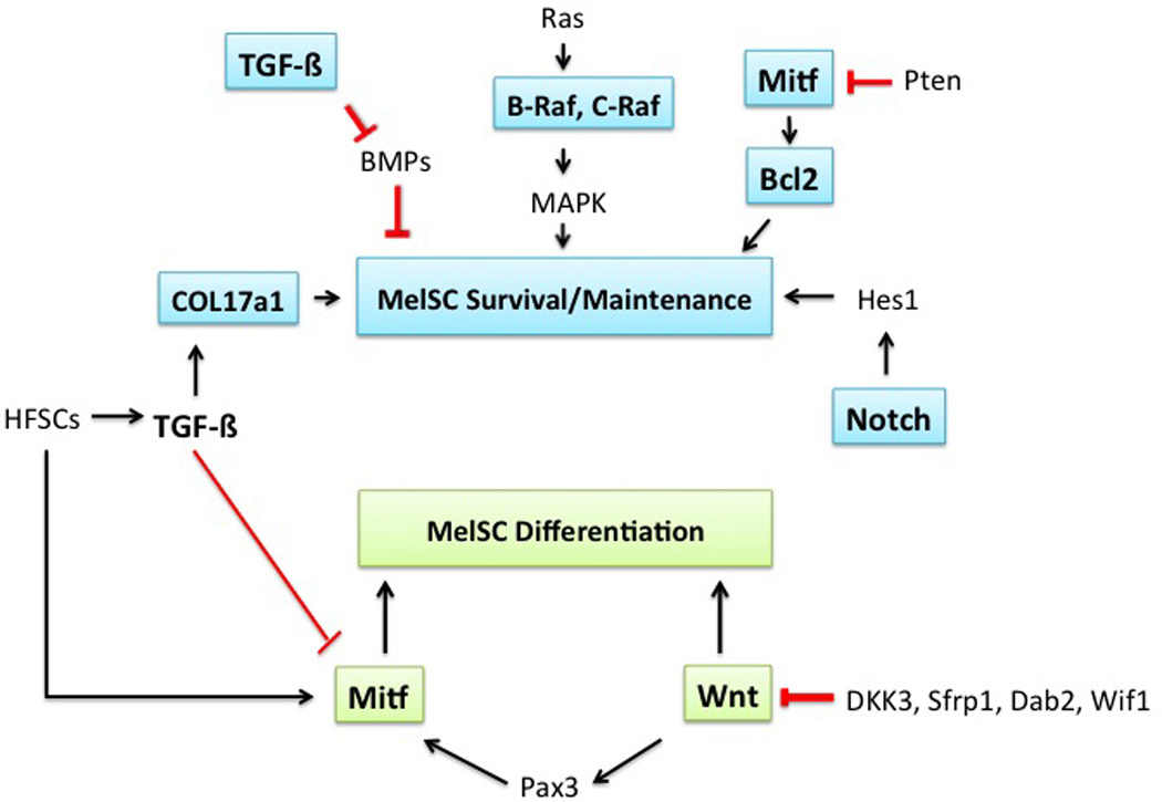
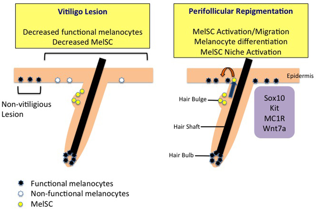
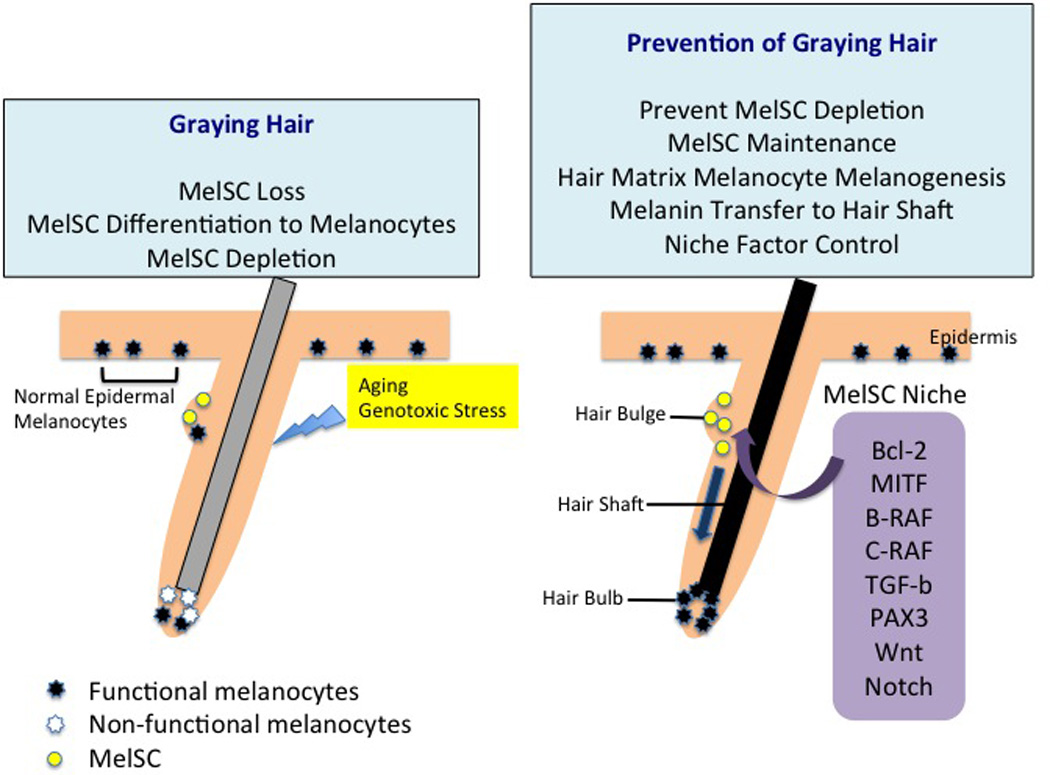
Introduction: Melanocytes produce pigment granules that color both skin and hair. In the hair follicles melanocytes are derived from stem cells (MelSCs) that are present in hair bulges or sub-bulge regions and function as melanocyte reservoirs. Quiescence, maintenance, activation and proliferation of MelSCs are controlled by specific activities in the microenvironment that can influence the differentiation and regeneration of melanocytes. Therefore, understanding MelSCs and their niche may lead to use of MelSCs in new treatments for various pigmentation disorders.
Areas covered: We describe here pathophysiological mechanisms by which melanocyte defects lead to skin pigmentation disorders such as vitiligo and hair graying. The development, migration and proliferation of melanocytes and factors involved in the survival, maintenance and regeneration of MelSCs are reviewed with regard to the biological roles and potential therapeutic applications in skin pigmentation diseases.
Expert opinion: MelSC biology and niche factors have been studied mainly in murine experimental models. Human MelSC markers or methods to isolate them are much less well understood. Identification, isolation and culturing of human MelSCs would represent a major step toward new biological therapeutic options for patients with recalcitrant pigmentary disorders or hair graying. By modulating the niche factors for MelSCs, it may one day be possible to control skin pigmentary disorders and prevent or reverse hair graying.
Keywords: graying hair; melanocyte; melanocyte stem cell; pigmentation; vitiligo.
Conflict of interest statement
The author has no relevant affiliations or financial involvement with any organization or entity with a financial interest in or financial conflict with the subject matter or materials discussed in the manuscript. This includes employment, consultancies, honoraria, stock ownership or options, expert testimony, grants or patents received or pending, or royalties.
Figures

Similar articles
-
Melanocyte stem cells: a melanocyte reservoir in hair follicles for hair and skin pigmentation.Pigment Cell Melanoma Res. 2011 Jun;24(3):401-10. doi: 10.1111/j.1755-148X.2011.00855.x. Epub 2011 May 5. Pigment Cell Melanoma Res. 2011. PMID: 21466661 Review.
-
Melanocyte Chitosan/Gelatin Composite Fabrication with Human Outer Root Sheath-Derived Cells to Produce Pigment.Sci Rep. 2019 Mar 26;9(1):5198. doi: 10.1038/s41598-019-41611-5. Sci Rep. 2019. PMID: 30914712 Free PMC article.
-
Lenalidomide Augments Differentiation of Cultured Hair Follicle Derived Melanocyte Stem Cells Into Functional Melanocytes.Dermatol Pract Concept. 2023 Apr 1;13(2):e2023077. doi: 10.5826/dpc.1302a77. Online ahead of print. Dermatol Pract Concept. 2023. PMID: 37196305 Free PMC article.
-
Skin pigmentation and its control: From ultraviolet radiation to stem cells.Exp Dermatol. 2021 Apr;30(4):560-571. doi: 10.1111/exd.14260. Epub 2020 Dec 24. Exp Dermatol. 2021. PMID: 33320376 Free PMC article. Review.
-
Melanocyte stem cells in skin diseases and their potential in cell-based therapy.Histol Histopathol. 2022 Oct;37(10):937-953. doi: 10.14670/HH-18-470. Epub 2022 May 13. Histol Histopathol. 2022. PMID: 35553404 Review.
Cited by
-
Melanocytes in regenerative medicine applications and disease modeling.J Transl Med. 2024 Apr 8;22(1):336. doi: 10.1186/s12967-024-05113-x. J Transl Med. 2024. PMID: 38589876 Free PMC article. Review.
-
Pigmented Epithelioid Melanocytoma (PEM)/Animal Type Melanoma (ATM): Quest for an Origin. Report of One Unusual Case Indicating Follicular Origin and Another Arising in an Intradermal Nevus.Int J Mol Sci. 2017 Aug 15;18(8):1769. doi: 10.3390/ijms18081769. Int J Mol Sci. 2017. PMID: 28809777 Free PMC article.
-
Three Streams for the Mechanism of Hair Graying.Ann Dermatol. 2018 Aug;30(4):397-401. doi: 10.5021/ad.2018.30.4.397. Epub 2018 Jun 28. Ann Dermatol. 2018. PMID: 30065578 Free PMC article. Review.
-
Autologous lipoaspirate as a new treatment of vulvar lichen sclerosus: A review on literature.Exp Dermatol. 2022 May;31(5):689-699. doi: 10.1111/exd.14561. Epub 2022 Mar 16. Exp Dermatol. 2022. PMID: 35276020 Free PMC article. Review.
-
Focus on the Contribution of Oxidative Stress in Skin Aging.Antioxidants (Basel). 2022 Jun 6;11(6):1121. doi: 10.3390/antiox11061121. Antioxidants (Basel). 2022. PMID: 35740018 Free PMC article. Review.
References
-
- Dupin E, Sommer L. Neural crest progenitors and stem cells: from early development to adulthood. Dev Biol. 2012;366(1):83–95. - PubMed
-
- Yaar M, Park HY. Melanocytes: a window into the nervous system. J Invest Dermatol. 2012;132(3 Pt 2):835–845. - PubMed
-
- Wittgen HG, van Kempen LC. Reactive oxygen species in melanoma and its therapeutic implications. Melanoma Res. 2007;17(6):400–409. - PubMed
-
- Funasaka Y, Komoto M, Ichihashi M. Depigmenting effect of alpha-tocopheryl ferulate on normal human melanocytes. Pigment Cell Res. 2000;13(Suppl 8):170–174. - PubMed
Publication types
MeSH terms
Grants and funding
LinkOut - more resources
Full Text Sources
Other Literature Sources
Medical