. 2014 Sep 19;289(38):26533-26541.
doi: 10.1074/jbc.M114.593970. Epub 2014 Aug 7.

Tyr-301 phosphorylation inhibits pyruvate dehydrogenase by blocking substrate binding and promotes the Warburg effect

Affiliations

Tyr-301 phosphorylation inhibits pyruvate dehydrogenase by blocking substrate binding and promotes the Warburg effect

Jun Fan et al. J Biol Chem. .

Abstract

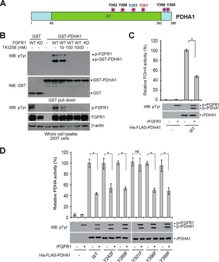
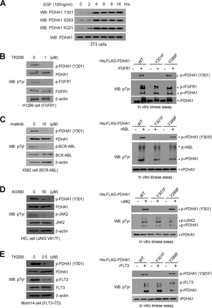
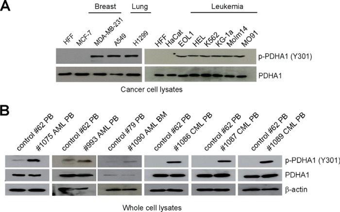
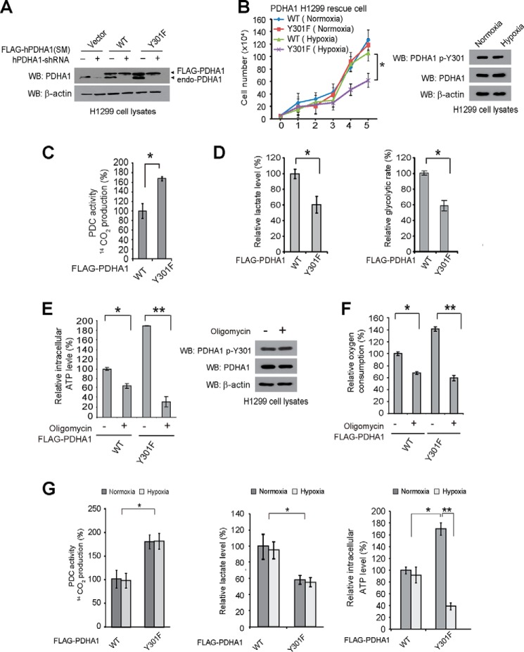
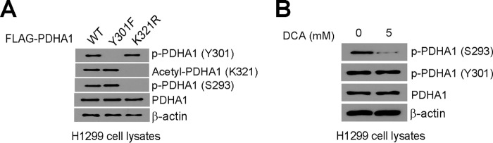
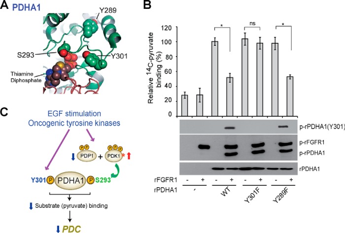
The mitochondrial pyruvate dehydrogenase complex (PDC) plays a crucial role in regulation of glucose homoeostasis in mammalian cells. PDC flux depends on catalytic activity of the most important enzyme component pyruvate dehydrogenase (PDH). PDH kinase inactivates PDC by phosphorylating PDH at specific serine residues, including Ser-293, whereas dephosphorylation of PDH by PDH phosphatase restores PDC activity. The current understanding suggests that Ser-293 phosphorylation of PDH impedes active site accessibility to its substrate pyruvate. Here, we report that phosphorylation of a tyrosine residue Tyr-301 also inhibits PDH α 1 (PDHA1) by blocking pyruvate binding through a novel mechanism in addition to Ser-293 phosphorylation. In addition, we found that multiple oncogenic tyrosine kinases directly phosphorylate PDHA1 at Tyr-301, and Tyr-301 phosphorylation of PDHA1 is common in EGF-stimulated cells as well as diverse human cancer cells and primary leukemia cells from human patients. Moreover, expression of a phosphorylation-deficient PDHA1 Y301F mutant in cancer cells resulted in increased oxidative phosphorylation, decreased cell proliferation under hypoxia, and reduced tumor growth in mice. Together, our findings suggest that phosphorylation at distinct serine and tyrosine residues inhibits PDHA1 through distinct mechanisms to impact active site accessibility, which act in concert to regulate PDC activity and promote the Warburg effect.

Keywords: Cell Proliferation; Phosphotyrosine Signaling; Pyruvate Dehydrogenase Complex (PDC); Tumor Metabolism; Warburg Effect.


Figures

FIGURE 1.
FIGURE 1.
FGFR1 inhibits PDHA1 via phosphorylation at Tyr-301. A, schematic representation of PDHA1. Five identified FGFR1 direct tyrosine phosphorylation sites are shown. B, Western blot (WB) detecting tyrosine phosphorylation of purified GST-PDHA1 from cells co-expressing FGFR1 WT or an FGFR1 kinase-dead form (KD) using a pan phosphotyrosine antibody. Cells treated with FGFR1 inhibitor TKI258 were incubated for 4 h at indicated concentrations. C, active rFGFR1 directly phosphorylates purified His-FLAG-PDHA1 at tyrosine residues in an in vitro kinase assay and attenuates PDHA1 activity. D, purified His-FLAG-PDHA1 variants were incubated with rFGFR1, followed by PDHA1 activity assay. The error bars represent mean values ± S.D. from three replicates of each sample (*, 0.01 < p < 0.05; **, 0.001 < p < 0.01; ns, not significant).
FIGURE 2.
FIGURE 2.
EGF stimulation and diverse oncogenic tyrosine kinases lead to Tyr-301 phosphorylation of PDHA1. A, 3T3 cells treated with EGF for increasing time were examined for phospho-PDHA1 (Tyr(P)-301 (pTyr301)) leve1 using immunoblotting. B–E, left panels: immunoblotting shows phosphorylation levels of PDHA1 Tyr-301 in FGFR1-expressing human lung cancer H1299 cells treated with FGFR1 inhibitor TKI258 (B), BCR-ABL-expressing leukemia K562 cells treated with ABL inhibitor imatinib (C), JAK2 V617F-expressing leukemia HEL cells treated with JAK2 inhibitor AG490 (D), or FLT3-ITD-expressing leukemia Molm 14 cells treated with FLT3 inhibitor TKI258 (E). Right panels: purified His-FLAG-PDHA1 WT, Y301F, or Y289F were incubated with rFGFR1 (B), rABL (C), rJAK2 (D), or rFLT3 (E), followed by immunoblotting to detect Tyr-301 phosphorylation of PDHA1. WB, Western blot.
FIGURE 3.
FIGURE 3.
Tyr-301 phosphorylation of PDHA1 is common in human cancer cells. A and B, immunoblotting to detect phosphorylation levels of PDHA1 Tyr-301 in diverse human tumor and leukemia cells (A) as well as human primary leukemia cells isolated from peripheral blood (PB) or bone marrow (BM) samples from representative acute myeloid leukemia (AML) and chronic myeloid leukemia (CML) patients (B). Normal proliferating human foreskin fibroblasts (HFF), HaCaT keratinocyte cells, and peripheral blood cells from healthy human donors were included as controls.
FIGURE 4.
FIGURE 4.
Expression of PDHA1 Y301F mutant in H1299 cells leads to decreased proliferation under hypoxia and increased oxidative phosphorylation. A, generation of H1299 cells with stable knockdown of endogenous hPDHA1, followed by stable rescue expression of FLAG-tagged hPDHA1 variants, which harbor silent mutations (SM) that confer PDHA1 shRNA-resistance. B, left: PDHA1 WT and Y301F rescue H1299 cells were tested for cell proliferation rate under normoxia (17% oxygen) or hypoxia (1% oxygen). Cell proliferation was determined based on cell numbers counted daily. Right: phosphorylation level of PDHA1 Tyr-301 was examined in H1299 cells under normoxic or hypoxic conditions using immunoblotting. C and D, PDHA1 WT or Y301F rescue H1299 cells were tested for PDC flux rate (C) as well as lactate production and glycolytic rate (D; left and right, respectively) under normoxia. E and F, PDHA1 rescue cells were tested for intracellular ATP level (E) and oxygen consumption (F) in the presence and absence of ATP synthase inhibitor oligomycin under normoxia. E, right: phosphorylation level of PDHA1 Tyr-301 was examined in H1299 cells in the presence or absence of oligomycin. G, PDHA1 rescue cells were tested for PDC flux rate (left), lactate production (middle), and intracellular ATP level (right) under normoxic or hypoxic conditions for 2 h. The error bars represent mean values ± S.D. from three replicates of each sample (*, 0.01 < p < 0.05; **, 0.001 < p < 0.01; ns, not significant). WB, Western blot.
FIGURE 5.
FIGURE 5.
Tyr-301 phosphorylation of PDHA1 is important for tumor growth. A and B, tumor growth (A) and (B) masses in xenograft nude mice injected with PDHA1 Y301F rescue cells compared with mice injected with control PDHA1 WT rescue cells are shown. p values were determined by a two-tailed paired Student's t test. C, top: dissected tumors in a representative nude mouse injected with PDHA1 WT and Y301F rescue cells on the left and right flanks, respectively, are shown. Bottom panel shows detection of Tyr-301 phosphorylation levels of PDHA1 in tumor lysates using specific phospho-PDHA1 (Tyr-301) antibody.
FIGURE 6.
FIGURE 6.
Tyr-301 phosphorylation provides a novel and distinct mechanism to inhibit PDHA1. A, distinct PDHA1 rescue cells were tested in immunoblotting to detect Tyr-301 phosphorylation, Lys-321 acetylation and Ser-293 phosphorylation levels. B, immunoblotting results detecting phosphorylation levels of Ser-293 and Tyr-301 in H1299 cells treated with PDK inhibitor dichloroacetate (DCA).
FIGURE 7.
FIGURE 7.
Tyr-301 phosphorylation inhibits PDHA1 by blocking substrate pyruvate binding. A, schematic representation of PDHA1 structure (Protein Data Bank code 1NI4 (32)). Tyr-301 is proximal to the PDHA1 catalytic site where substrate (pyruvate) binds (<10 Å), a distance similar to Ser-293 pyruvate. B, purified His-FLAG-PDHA1 variants were incubated with rFGFR1, followed by incubation with 2-14C-labeled pyruvate. PDHA1-bound [2-14C]pyruvate was assessed by scintillation counting. The error bars represent mean values ± S.D. from three replicates of each sample (*, 0.01 < p < 0.05; **, 0.001 < p < 0.01; ns, not significant). C, proposed model shows that phosphorylation at Tyr-301 inhibits PDHA1 and blocks substrate pyruvate binding through a novel and distinct mechanism in addition to Ser-293 phosphorylation in EGF-stimulated cells and cancer cells where tyrosine kinase signaling is commonly up-regulated.

References

    1. Cairns R. A., Harris I. S., Mak T. W. (2011) Regulation of cancer cell metabolism. Nat. Rev. Cancer 11, 85–95 - PubMed
    1. Kroemer G., Pouyssegur J. (2008) Tumor cell metabolism: cancer's Achilles' heel. Cancer Cell 13, 472–482 - PubMed
    1. Hiromasa Y., Fujisawa T., Aso Y., Roche T. E. (2004) Organization of the cores of the mammalian pyruvate dehydrogenase complex formed by E2 and E2 plus the E3-binding protein and their capacities to bind the E1 and E3 components. J. Biol. Chem. 279, 6921–6933 - PubMed
    1. Read R. J. (2001) Pushing the boundaries of molecular replacement with maximum likelihood. Acta Crystallogr. D 57, 1373–1382 - PubMed
    1. Harris R. A., Bowker-Kinley M. M., Huang B., Wu P. (2002) Regulation of the activity of the pyruvate dehydrogenase complex. Adv. Enzyme Regul. 42, 249–259 - PubMed

Publication types

MeSH terms

Substances

Associated data

LinkOut - more resources