. 2014 Aug;19(8):086007.
doi: 10.1117/1.JBO.19.8.086007.

Handheld photoacoustic tomography probe built using optical-fiber parallel acoustic delay lines

Affiliations

Handheld photoacoustic tomography probe built using optical-fiber parallel acoustic delay lines

Young Cho et al. J Biomed Opt. 2014 Aug.

Abstract

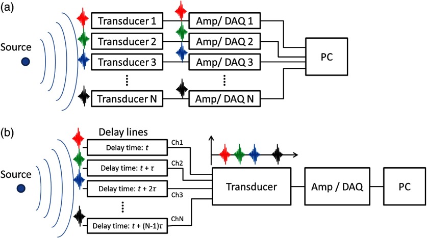
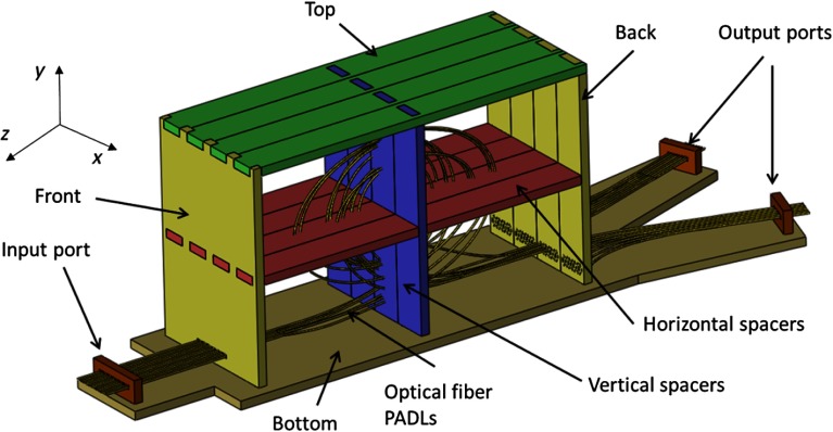
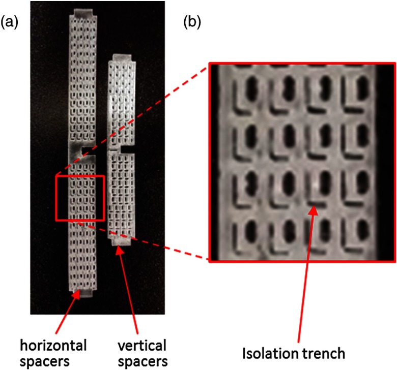
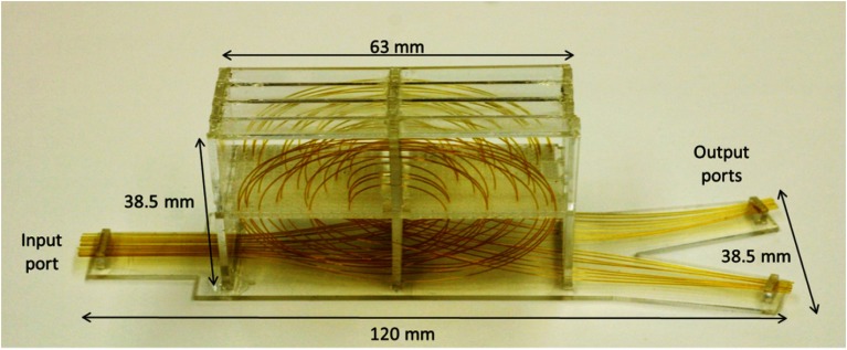
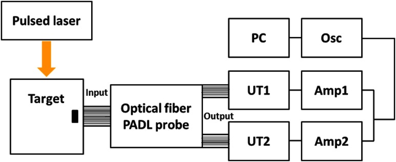
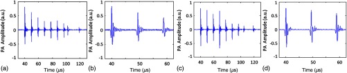
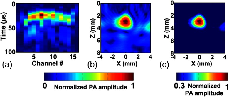
The development of the first miniaturized parallel acoustic delay line (PADL) probe for handheld photoacoustic tomography (PAT) is reported. Using fused-silica optical fibers with low acoustic attenuation, we constructed two arrays of eight PADLs. Precision laser micromachining was conducted to produce robust and accurate mechanical support and alignment structures for the PADLs, with minimal acoustic distortion and interchannel coupling. The 16 optical-fiber PADLs, each with a different time delay, were arranged to form one input port and two output ports. A handheld PADL probe was constructed using two single-element transducers and two data acquisition channels (equal to a channel reduction ratio of 8∶1). Photoacoustic (PA) images of a black-ink target embedded in an optically scattering phantom were successfully acquired. After traveling through the PADLs, the eight channels of differently time-delayed PA signals reached each single-element ultrasonic transducer in a designated nonoverlapping time series, allowing clear signal separation for PA image reconstruction. Our results show that the PADL technique and the handheld probe can potentially enable real-time PAT, while significantly reducing the complexity and cost of the ultrasound receiver system.


Figures

Fig. 1
Fig. 1
(a) Photoacoustic (PA) signal reception using multiple transducers and multichannel data acquisition (DAQ) electronics; and (b) PA signal reception using a single-element transducer and single-channel DAQ electronics through parallel acoustic delay lines.
Fig. 2
Fig. 2
Schematic of the 16-channel handheld parallel acoustic delay line (PADL) probe; a perspective view of the probe input port, the PADL housing unit, and two output ports.
Fig. 3
Fig. 3
(a) Horizontal and vertical spacers fabricated using laser micromachining; and (b) zoom-in view of the horizontal spacer showing the elliptical threading holes and “L” shaped isolation trenches.
Fig. 4
Fig. 4
Fully assembled optical fiber PADL probe.
Fig. 5
Fig. 5
Photoacoustic tomography imaging setup using the 16-channel PADL probe.
Fig. 6
Fig. 6
Plots of acoustic waveforms propagating through the optical-fiber PADLs with various lengths ranging from 21 to 56 cm: (a) Raw A-line signals obtained by ultrasonic transducer UT1; (b) zoom-in view of the first three signals in (a); (c) raw A-line image obtained by ultrasonic transducer UT2; and (d) zoom-in view of the first three signals in (c).
Fig. 7
Fig. 7
(a) Photograph of an optically absorbing target embedded in an optically scattering medium; (b) raw A-line PA signals with Hilbert transformation obtained by the ultrasonic transducer (UT 1); and (c) raw A-line PA signals with Hilbert transformation obtained by the ultrasonic transducer (UT 2).
Fig. 8
Fig. 8
(a) Rearranged Hilbert-transformed raw data acquired by 16 optical fibers; (b) reconstructed PA images with compensation for the acoustic attenuation in each fiber; and (c) thresholded (30%) PA image of (b).

References

    1. Oraevsky A. A., Karabutov A. A., Biomedical Photonics Handbook, Vol. PM125, CRC, Boca Raton, Florida: (2003).
    1. Kim C., Favazza C., Wang L. V., “In vivo photoacoustic tomography of chemicals: high-resolution functional and molecular optical imaging at new depths,” Chem. Rev. 110(5), 2756–2782 (2010).CHREAY10.1021/cr900266s - DOI - PMC - PubMed
    1. Wang L., Photoacoustic Imaging and Spectroscopy, Vol. 144, CRC, Boca Raton, Florida: (2009).
    1. Wang L. V., Hu S., “Photoacoustic tomography: in vivo imaging from organelles to organs,” Science 335(6075), 1458–1462 (2012).SCIEAS10.1126/science.1216210 - DOI - PMC - PubMed
    1. Fronheiser M. P., et al. , “Real-time optoacoustic monitoring and three-dimensional mapping of a human arm vasculature,” J. Biomed. Opt. 15(2), 021305 (2010).JBOPFO10.1117/1.3370336 - DOI - PMC - PubMed

Publication types

MeSH terms