Review
. 2014 Aug 7;6(9):a016428.
doi: 10.1101/cshperspect.a016428.

Sources of DNA double-strand breaks and models of recombinational DNA repair

Affiliations
Review

Sources of DNA double-strand breaks and models of recombinational DNA repair

Anuja Mehta et al. Cold Spring Harb Perspect Biol. .

Abstract

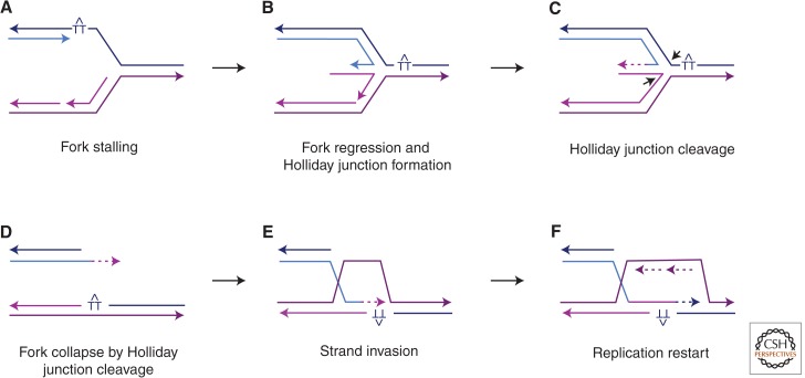
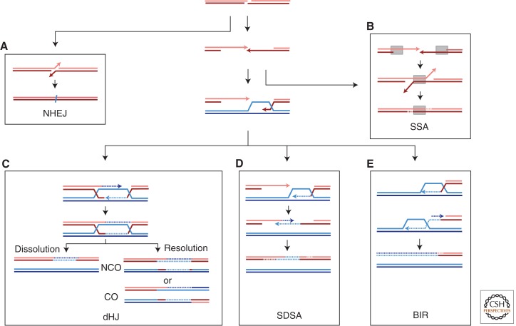
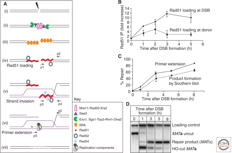
DNA is subject to many endogenous and exogenous insults that impair DNA replication and proper chromosome segregation. DNA double-strand breaks (DSBs) are one of the most toxic of these lesions and must be repaired to preserve chromosomal integrity. Eukaryotes are equipped with several different, but related, repair mechanisms involving homologous recombination, including single-strand annealing, gene conversion, and break-induced replication. In this review, we highlight the chief sources of DSBs and crucial requirements for each of these repair processes, as well as the methods to identify and study intermediate steps in DSB repair by homologous recombination.


Figures

Figure 1.
Figure 1.
Replication fork stalling and restart. DNA replication can be stalled at UV-induced thymidine dimers (TT), as well as DNA secondary structures. A stalled replication fork (A) can undergo regression and pairing of the newly synthesized strands to form a HJ “chicken foot” intermediate (B). The HJ can be cleaved by HJ resolvases (C) to lead to a collapsed fork, effectively a one-ended chromosome break (D). The free end can initiate HR by strand invasion (E) to bypass the lesion and resume replication (F). Newly synthesized DNA is depicted as dashed lines in the same color as the template; arrowheads indicate 3′ ends.
Figure 2.
Figure 2.
Pathways of DNA DSB repair. DSBs are efficiently repaired by HR and NHEJ. DSBs are processed by 5′ to 3′ end resection producing 3′ single-stranded tails. (A) NHEJ involves ligation of broken ends, with little or no base pairing, to produce small deletions or insertions. (B) Single-strand annealing (SSA) takes place when resection reveals flanking homologous repeats that can anneal, leading to deletion of the intervening sequences. (C,D) Repair by two different mechanisms of GC results in a short patch of new DNA synthesis to repair the DSB. (C) In synthesis-dependent strand-annealing (SDSA), the newly synthesized strand dissociates from the D-loop and results in a noncrossover (NCO) outcome with no change to the template DNA. (D) The double Holliday Junction (dHJ) pathway involves second end capture to stabilize the D-loop. The dHJ structure can be resolved either by helicase and topoisomerase-mediated dissolution to give NCO or cleaved by HJ resolvases to produce both crossover (CO) or NCO outcomes. (E) BIR involves both leading and lagging strand synthesis and results in loss of heterozygosity or, if the template is located ectopically, a nonreciprocal translocation. Newly synthesized DNA is depicted as dashed lines in the same color as the template; arrowheads indicate 3′ ends.
Figure 3.
Figure 3.
Key intermediate steps of HR and methods to study them. (A) Key proteins are depicted in sequential early steps in GC in budding yeast. DSB formation (i) is immediately followed by 5′ to 3′ resection (ii). The 3′ tails are stabilized by RPA (iii), which is then replaced with Rad51 recombinase with assistance from accessory proteins like Rad52 (iv). Once the homologous donor is found, strand invasion occurs, resulting in formation of a D-loop (v) by displacement of the identical strand and base pairing with the complementary strand at the donor. (vi) Various components of the replication machinery assemble to start copying from the donor template. (vii) The break is sealed. Small arrows denote positions of PCR primer pairs used to analyze intermediate steps, shown on the right for MAT switching in S. cerevisiae. (B) Recruitment of Rad51 at DSB site (the MATa locus) by chromatin immunoprecipitation (ChIP) followed by quantitative PCR (qPCR) using primers p1 and p2 (solid line). Rad51 binding to the donor template (budding yeast HMLα locus) using primers p3 and p4 for qPCR (dotted line). Error bars indicate standard error of the mean. (C) The initiation of new DNA synthesis by primer extension is detected by using PCR primers p5 and p6, which amplifies a unique fragment once new DNA synthesis has been initiated (solid line). A dotted line shows quantitative densitometric analysis of Southern blot (D) that follows GC progression in budding yeast as MATa switches to MATα. Data from the investigators (A Mehta and J Haber, unpubl.).

References

    1. Aguilera A, Gaillard H 2014. Transcription and recombination: When RNA meets DNA. Cold Spring Harb Perspect Biol 10.1101/cshperspect.a016543 - DOI - PMC - PubMed
    1. Arcangioli B 2000. Fate of mat1 DNA strands during mating-type switching in fission yeast. EMBO Rep 1: 145–150 - PMC - PubMed
    1. Aylon Y, Liefshitz B, Kupiec M 2004. The CDK regulates repair of double-strand breaks by homologous recombination during the cell cycle. EMBO J 23: 4868–4875 - PMC - PubMed
    1. Balestrini A, Ristic D, Dionne I, Liu XZ, Wyman C, Wellinger RJ, Petrini JH 2013. The Ku heterodimer and the metabolism of single-ended DNA double-strand breaks. Cell Rep 3: 2033–2045 - PMC - PubMed
    1. Barlow JH, Lisby M, Rothstein R 2008. Differential regulation of the cellular response to DNA double-strand breaks in G1. Mol Cell 30: 73–85 - PMC - PubMed

Publication types