. 2014 Aug 7;95(2):227-34.
doi: 10.1016/j.ajhg.2014.07.007.

Mutations in LAMA1 cause cerebellar dysplasia and cysts with and without retinal dystrophy

Collaborators, Affiliations

Mutations in LAMA1 cause cerebellar dysplasia and cysts with and without retinal dystrophy

Kimberly A Aldinger et al. Am J Hum Genet. .

Erratum in

  • Am J Hum Genet. 2014 Oct 2;95(4):472

Abstract

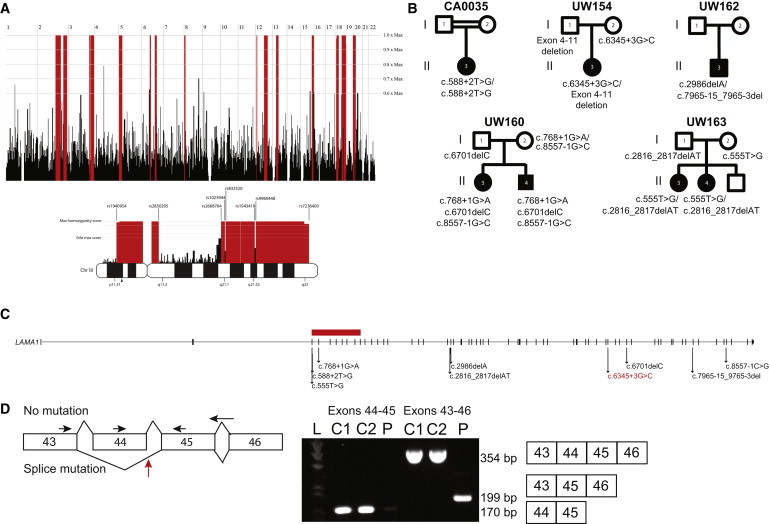
Cerebellar dysplasia with cysts (CDC) is an imaging finding typically seen in combination with cobblestone cortex and congenital muscular dystrophy in individuals with dystroglycanopathies. More recently, CDC was reported in seven children without neuromuscular involvement (Poretti-Boltshauser syndrome). Using a combination of homozygosity mapping and whole-exome sequencing, we identified biallelic mutations in LAMA1 as the cause of CDC in seven affected individuals (from five families) independent from those included in the phenotypic description of Poretti-Boltshauser syndrome. Most of these individuals also have high myopia, and some have retinal dystrophy and patchy increased T2-weighted fluid-attenuated inversion recovery (T2/FLAIR) signal in cortical white matter. In one additional family, we identified two siblings who have truncating LAMA1 mutations in combination with retinal dystrophy and mild cerebellar dysplasia without cysts, indicating that cysts are not an obligate feature associated with loss of LAMA1 function. This work expands the phenotypic spectrum associated with the lamininopathy disorders and highlights the tissue-specific roles played by different laminin-encoding genes.


Figures

Figure 1
Figure 1
Mutation Identification in Individuals with CDC (A) Homozygosity mapping in CA0035 identified several regions of homozygosity (red) across the genome. Homozygous regions on chromosome 18 are indicated below the genome-wide mapping. An arrowhead below the chromosome 18 ideogram indicates the location of LAMA1. (B) Pedigree diagrams showing segregation analysis for five families. (C) LAMA1 is composed of 63 exons that span chr18: 6,941,743–7,117,813 (UCSC Genome Browser hg19) and encode protein-coding sequence. Arrows indicate the locations of ten different mutations identified in five families. The horizontal red bar represents a deletion spanning LAMA1 exons 4–11. (D) The splice-site mutation (designated red in C) in UW154-3 results in skipping of exon 45. Total RNA was isolated from fibroblasts with the RNeasy Mini Kit (QIAGEN) and transcribed into cDNA with the SuperScript III First-Strand System and random hexamer primers (LifeTechnologies). RT-PCR was performed under standard conditions with forward primers in exons 43 and 44 and reverse primers in exons 45 and 46 (sequences are available upon request). Primers in exons 44 and 45 generated a 170 bp amplicon in cDNA from two unrelated, unaffected control fibroblasts but minimal product in cDNA from UW154-3. Primers in exons 43 and 46 generated a 354 bp amplicon in control cells and a 199 bp product in UW154-3. Sanger sequencing of this product revealed that exon 43 is spliced to exon 45 in UW154-3 (data not shown). The horizontal arrows indicate primer positions. The red arrow indicates the location of the splice-site mutation in intron 44. “L” indicates the ladder lane, “C1” and “C2” indicate the two control samples, and “P” indicates individual UW154-3. The expected size and exon composition of the respective products are indicated to the right of the gel.
Figure 2
Figure 2
Brain and Eye Imaging Findings in Affected Individuals with LAMA1 Mutations (A–E) Individual CA0035 has cerebellar cysts (arrowheads in A), superior cerebellar dysplasia (arrow in B) and large globes (B), elevated and splayed superior cerebellar peduncles (arrowheads in C), an enlarged fourth ventricle (A and D), and patchy increased T2/FLAIR (asterisks in E). The periventricular heterotopia (arrowhead in E) is atypical. (F–J) Similar findings in UW154-3 include cerebellar cysts (arrowhead in F), superior cerebellar dysplasia (arrowhead in G) and large globes (G), elevated and splayed superior cerebellar peduncles (arrowheads in H), an enlarged fourth ventricle (F and I), and patchy increased T2/FLAIR (asterisks in E). In addition, UW154-3 has partial absence of the corpus callosum (bracket in I) and septum pellucidum (plus sign in J). (K–N) UW163-3 has only superior cerebellar dysplasia (arrow in L) and patchy increased T2/FLAIR (asterisks in O), but no cerebellar cysts (K). The superior cerebellar peduncles (M) and the fourth ventricle (K and N) are normal.

References

    1. Demaerel P. Abnormalities of cerebellar foliation and fissuration: classification, neurogenetics and clinicoradiological correlations. Neuroradiology. 2002;44:639–646. - PubMed
    1. Jissendi-Tchofo P., Pandit F., Soto-Ares G., Vallee L. Neuropsychological evaluation and follow-up of children with cerebellar cortical dysplasia. Dev. Med. Child Neurol. 2011;53:1119–1127. - PubMed
    1. Poretti A., Boltshauser E. Cerebellar dysplasia. In: Boltshauser E., Schmahmann J., editors. Cerebellar disorders in children. Mac Keith Press; London: 2012. pp. 172–176.
    1. Soto-Ares G., Delmaire C., Deries B., Vallee L., Pruvo J.P. Cerebellar cortical dysplasia: MR findings in a complex entity. AJNR Am. J. Neuroradiol. 2000;21:1511–1519. - PMC - PubMed
    1. Boddaert N., Desguerre I., Bahi-Buisson N., Romano S., Valayannopoulos V., Saillour Y., Seidenwurm D., Grevent D., Berteloot L., Lebre A.S. Posterior fossa imaging in 158 children with ataxia. J. Neuroradiol. 2010;37:220–230. - PubMed

Publication types

LinkOut - more resources