
Endothelial C-type natriuretic peptide maintains vascular homeostasis

Amie J Moyes et al. J Clin Invest. 2014 Sep.

Abstract

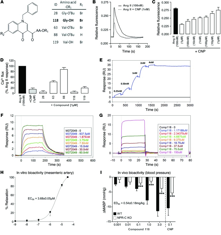
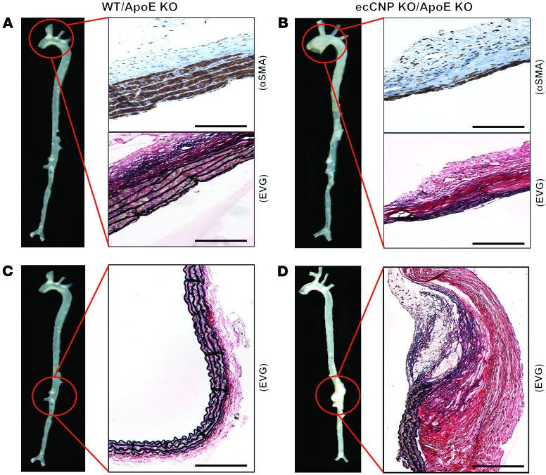
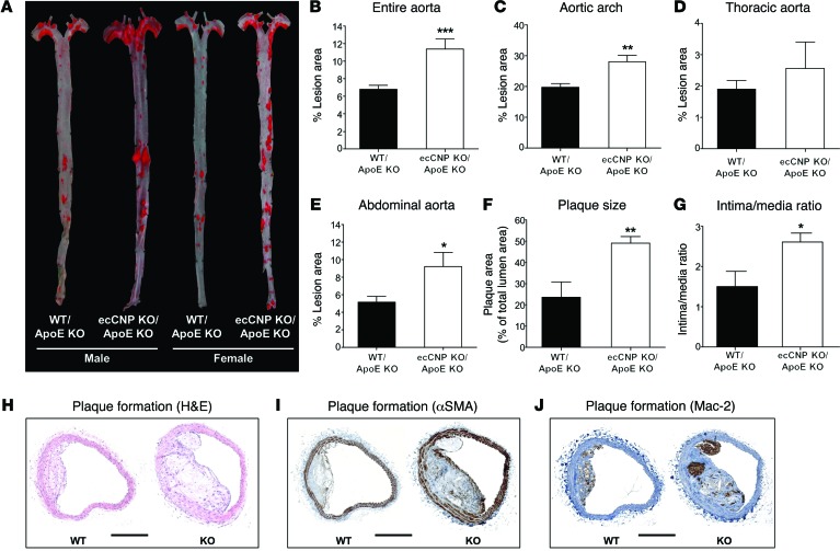
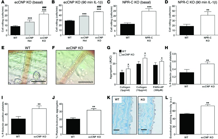
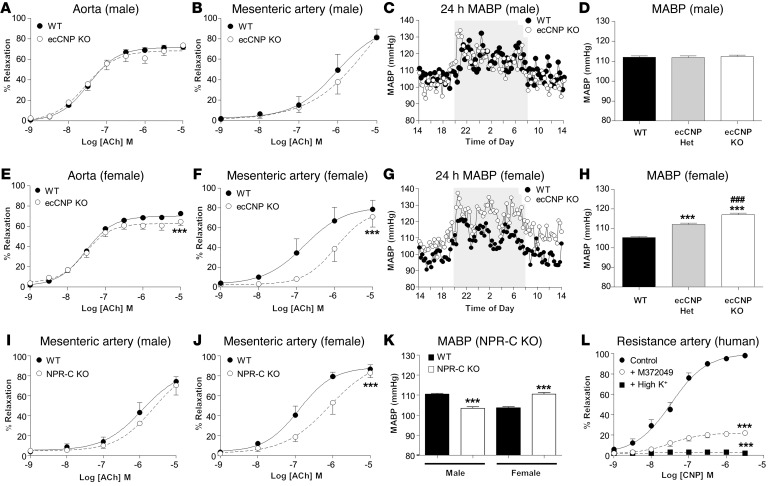
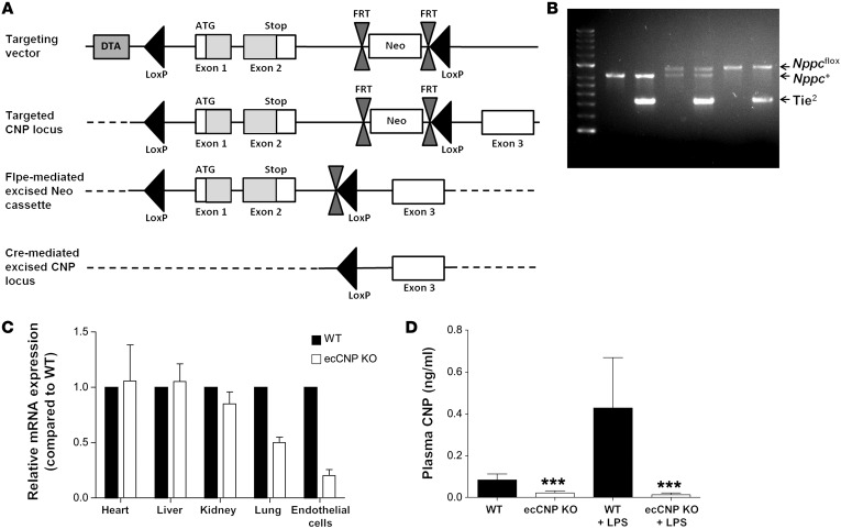
The endothelium plays a fundamental role in maintaining vascular homeostasis by releasing factors that regulate local blood flow, systemic blood pressure, and the reactivity of leukocytes and platelets. Accordingly, endothelial dysfunction underpins many cardiovascular diseases, including hypertension, myocardial infarction, and stroke. Herein, we evaluated mice with endothelial-specific deletion of Nppc, which encodes C-type natriuretic peptide (CNP), and determined that this mediator is essential for multiple aspects of vascular regulation. Specifically, disruption of CNP leads to endothelial dysfunction, hypertension, atherogenesis, and aneurysm. Moreover, we identified natriuretic peptide receptor-C (NPR-C) as the cognate receptor that primarily underlies CNP-dependent vasoprotective functions and developed small-molecule NPR-C agonists to target this pathway. Administration of NPR-C agonists promotes a vasorelaxation of isolated resistance arteries and a reduction in blood pressure in wild-type animals that is diminished in mice lacking NPR-C. This work provides a mechanistic explanation for genome-wide association studies that have linked the NPR-C (Npr3) locus with hypertension by demonstrating the importance of CNP/NPR-C signaling in preserving vascular homoeostasis. Furthermore, these results suggest that the CNP/NPR-C pathway has potential as a disease-modifying therapeutic target for cardiovascular disorders.


Figures

Figure 6
Figure 6. Chemical and biological characterization of NPR-C agonists.
Structure of selected NPR-C agonists (A). Representative trace showing inhibition of the Ca2+ flux in response to angiotensin II (Ang II; 100 nM) by CNP in primary mesenteric artery smooth muscle cells using a FLIPR-based assay (B and C). Structure-activity relationship (SAR) for selected NPR-C agonist series using an identical cell-based assay (D). SPR spectroscopic analysis of the interaction of CNP (0.25–4 nM) (E), M372049 (1–60 nM) (F), and compound 118 (1–150 μM) (G) with human NPR-C (representative of 3 separate experiments). Vasorelaxant activity of lead NPR-C agonist, compound 118, in isolated mesenteric arteries (H), and the hypotensive effect of compound 118 in WT and NPR-C KO mice (I). Data are represented as the mean ± SEM. n = 6 for the in vitro and in vivo vascular reactivity studies and cell-based assays. ***P < 0.001, significantly different from WT littermates.
Figure 5
Figure 5. Aneurysm development in ecCNP KO mice.
Images of the aneurysms in male ecCNP KO/ApoE KO mice show dilation of the aortic arch (B) or abdominal aorta (D), loss of vascular smooth muscle and thinning of the vascular wall (B), and degradation of elastin (D) in comparison with WT/ApoE KO animals (A and C). Scale bars: 50 μm. Representative of 4/9 male animals. EVG, elastic van Gieson.
Figure 4
Figure 4. Accelerated atherogenesis in ecCNP KO mice.
Atherosclerotic plaque formation was accelerated in the ecCNP/ApoE dKO animals in comparison with WT/ApoE KO. A representative image of oil red O staining of the lesions in the aortic tree is shown (A), with quantification of plaque area in the entire aorta (B), aortic arch (C), thoracic aorta (D), and abdominal aorta (E). Histological staining of the brachiocephalic arteries from these mice showed a larger plaque size (F and H), greater intima media thickness ratio (G and I), and macrophage infiltration (J). Data are represented as the mean ± SEM. n = 18. *P < 0.05, **P < 0.01, ***P < 0.001, significantly different from WT/ApoE KO littermates. Scale bars: 200 μm (HJ).
Figure 3
Figure 3. Leukocyte and platelet hyperreactivity in ecCNP KO mice.
Basal and IL-1β–stimulated (5 ng/mouse; i.p.) leukocyte rolling was significantly greater in ecCNP KO (A and B) and NPR-C KO (C and D) mice compared with WT littermates. Representative images of basal rolling are shown (E and F). Platelet aggregation in response to collagen (3 μg/ml and 10 μg/ml) and PAR4-AP (300 μM; G), basal and PAR4-AP–triggered platelet expression of P-selectin (H and I), and the level of circulating platelet-leukocyte aggregates (J) was greater in ecCNP KO mice compared with WT littermates, implying that CNP regulates platelet function (and leukocyte recruitment) via inhibition of P-selectin expression. Endothelial P-selectin expression was also significantly greater in the aorta of ecCNP KO versus WT mice (K and L). Data are represented as the mean ± SEM. n = 10 for leukocyte studies; n = 6 for platelet reactivity and flow cytometry experiments. *P < 0.05, **P < 0.01, ***P < 0.001, significantly different from WT littermates; ###P < 0.001, significantly different from heterozygote littermates. Scale bars: 50 μm (E and F); 20 μm (K).
Figure 2
Figure 2. Endothelial dysfunction and hypertension in ecCNP KO mice.
Endothelium-dependent relaxation to ACh in isolated aorta and mesenteric artery of male ecCNP KO animals was unaltered in comparison with that of littermate controls (A and B). In contrast, there was a small reduction in maximal ACh relaxation in the aorta (E) and significant decrease in potency to ACh in the mesenteric arteries (F) of female ecCNP KO animals. MABP of male (C and D) and female (G and H) WT and ecCNP KO animals paralleled the in vitro vascular reactivity with a hypertensive phenotype only apparent in females; heterozygous ecCNP animals exhibited an intermediate phenotype (H). An analogous endothelial dysfunction (J) and hypertensive phenotype (K) was apparent in female NPR-C KO animals, whereas male NPR-C KO mice maintained normal endothelial function (I) and exhibited a mild hypotensive phenotype (K). CNP relaxed human mesenteric resistance arteries in a concentration-dependent manner and was sensitive to the NPR-C antagonist M372049 and high [K+] (L). Vasorelaxant responses in B, F, I, J, and L were obtained in the presence of L-NAME and indomethacin. Data are represented as the mean ± SEM. n = 6 for isolated vessel studies, n = 8 for the in vivo MABP studies. ***P < 0.001, significantly different from WT littermates; ###P < 0.01, significantly different from heterozygote littermates.
Figure 1
Figure 1. Development and characterization of an endothelium-specific CNP knockout mouse.
Cell-specific deletion of the CNP gene was achieved by flanking exons 1 and 2 of the CNP (Nppc) gene with LoxP sites (A). Flippase recognition target (FRT) sites were used to permit efficient removal of the neomycin cassette by breeding chimeric mice with ubiquitous Flpe-expressing animals, resulting in the generation of Nppc+/fl offspring. Nppcfl/fl animals were then crossed with a mouse in which expression of Cre recombinase is driven by an endothelial-specific promoter/enhancer associated with the angiopoietin Tie2 receptor. Heterozygous animals at the Nppc locus that expressed the Tie2 transgene (Tie2-Cre Nppc+/fl) were used as breeding pairs to generate ecCNP KO and corresponding WT (Tie2-Cre Nppc+/+) littermate controls. Analysis of the DNA from these animals confirmed generation of the 6 possible genotypes (B). qPCR analysis of CNP mRNA from different tissues established that the CNP gene had been deleted selectively from vascular endothelial cells (C). Measurement of plasma CNP concentrations under basal conditions and following administration of LPS (12.5 mg/kg; i.p.; 12 h) confirmed that peptide levels were significantly reduced in ecCNP KO versus WT animals (D). Data are represented as the mean ± SEM. n = 5. ***P < 0.001, significantly different from corresponding WT littermates.

References

    1. Vane JR, Anggard EE, Botting RM. Regulatory functions of the vascular endothelium. N Engl J Med. 1990;323(1):27–36. doi: 10.1056/NEJM199007053230106. - DOI - PubMed
    1. Moncada S, Higgs A. The L-arginine-nitric oxide pathway. N Engl J Med. 1993;329(27):2002–2012. doi: 10.1056/NEJM199312303292706. - DOI - PubMed
    1. Celermajer DS, Sorensen KE, Bull C, Robinson J, Deanfield JE. Endothelium-dependent dilation in the systemic arteries of asymptomatic subjects relates to coronary risk factors and their interaction. J Am Coll Cardiol. 1994;24(6):1468–1474. doi: 10.1016/0735-1097(94)90141-4. - DOI - PubMed
    1. Widlansky ME, Gokce N, Keaney JF, Keaney JF, Jr, Vita JA. The clinical implications of endothelial dysfunction. J Am Coll Cardiol. 2003;42(7):1149–1160. doi: 10.1016/S0735-1097(03)00994-X. - DOI - PubMed
    1. Ignarro LJ, Buga GM, Wood KS, Byrns RE, Chaudhuri G. Endothelium-derived relaxing factor produced and released from artery and vein is nitric oxide. Proc Natl Acad Sci U S A. 1987;84(24):9265–9269. doi: 10.1073/pnas.84.24.9265. - DOI - PMC - PubMed

Publication types