Review
. 2014 Aug 7;15(2):123-38.
doi: 10.1016/j.stem.2014.07.012.

Repair and regeneration of the respiratory system: complexity, plasticity, and mechanisms of lung stem cell function

Affiliations
Review

Repair and regeneration of the respiratory system: complexity, plasticity, and mechanisms of lung stem cell function

Brigid L M Hogan et al. Cell Stem Cell. .

Abstract

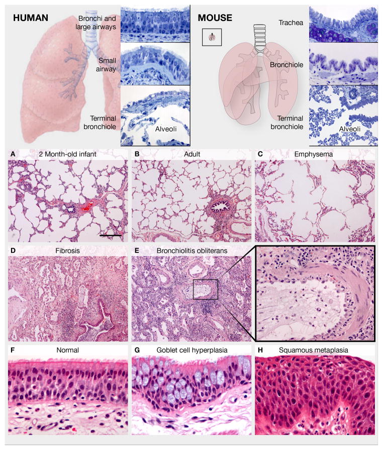
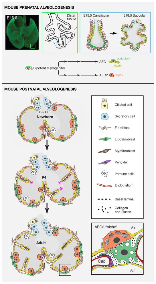
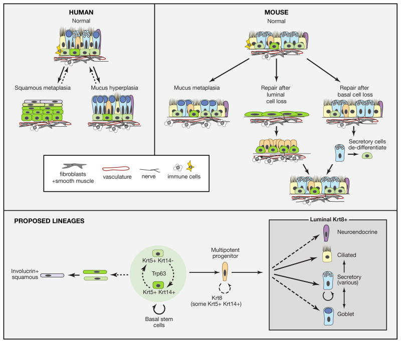
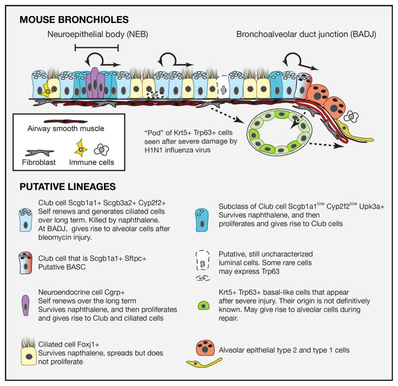
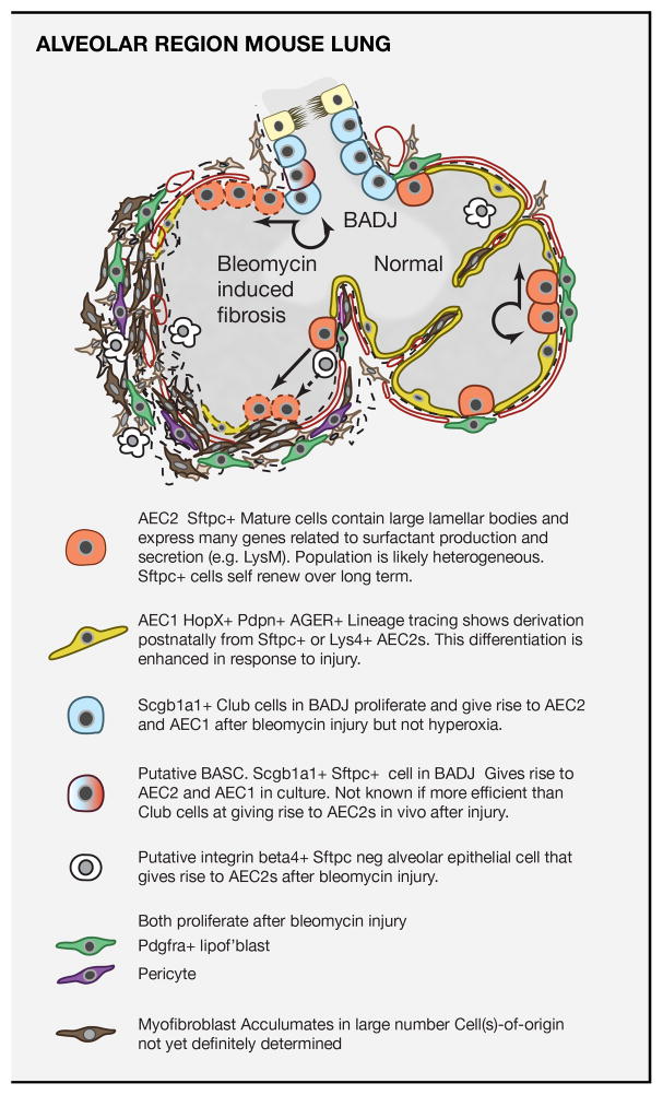
Respiratory disease is the third leading cause of death in the industrialized world. Consequently, the trachea, lungs, and cardiopulmonary vasculature have been the focus of extensive investigations. Recent studies have provided new information about the mechanisms driving lung development and differentiation. However, there is still much to learn about the ability of the adult respiratory system to undergo repair and to replace cells lost in response to injury and disease. This Review highlights the multiple stem/progenitor populations in different regions of the adult lung, the plasticity of their behavior in injury models, and molecular pathways that support homeostasis and repair.


Figures

Figure 1
Figure 1. Anatomy of the adult human and mouse lung and examples of human lung pathology
Upper panels: Regional epithelial histology in human and mouse. Left panel: The human trachea, bronchi and bronchioles >1 mm in diameter are lined by a pseudostratified epithelium with basal, multiciliated and secretory cells. Mucous goblet cells predominate in the larger airways, and Club cells in the smaller airways. Individual neuroendocrine cells and neuroendocrine bodies (NEBs) are scattered in the larger airways and increase distally. Cartilage, smooth muscle and stromal cells are associated with intralobar airways down to the small bronchioles. The simple cuboidal epithelium lining the terminal bronchioles leading into the alveoli is poorly characterized. The alveoli are lined by squamous AEC1s and cuboidal AEC2s. Right panel: In the mouse, only the trachea and mainstem bronchi have cartilage and a pseudostratified mucociliary epithelium with basal cells. The smaller bronchi and bronchioles are lined by a simple epithelium with multiciliated and Club cells, and fewer neuroendocrine cells and NEBs. The inset illustrates a mouse lung to the same scale as the human lung in left panel. Lower panels: Normal and pathologic human lung. (A, B) Images of the alveolar region in a 2 month-old infant and a normal adult illustrate that alveolar number increases postnatally through secondary alveolar septal crest formation. (C) In pulmonary emphysema, septal destruction and loss of alveolar cells results in alveolar enlargement. (D) In pulmonary fibrosis, the terminal bronchioles are plugged with mucus, alveolar epithelial morphology is abnormal, and alveolar architecture is dramatically altered by fibroblastic deposition of extracellular matrix. (E, E′) Bronchiolitis obliterans syndrome showing massive infiltration of immune cells, severe disruption of the small airway epithelium, and thickening of the underlying smooth muscle and stroma (boxed region magnified in E′). (F) Normal pseudostratified mucociliary bronchial epithelium from a lung transplant donor. (G, H) Goblet secretory cell hyperplasia and squamous metaplasia, respectively, in chronic obstructive lung disease. A–E and F–H, respectively, are the same magnification. Scale bar in A is 400 um.
Figure 2
Figure 2. Pre- and postnatal alveologenesis in the mouse
Upper panel: Schematic of the canalicular and saccular stages indicating that precursors of alveolar epithelial cells are laid down before birth. Image of whole mount E16 lung immunostained for e-cadherin provided by Ross Metzger. A distal canalicular tubule adjacent to the outer mesothelium is shown in greater detail (boxes). Evidence supports a model in which the population of distal tip cells (that are Sox9+ Id2+) is bipotential, expresses markers of both mature AEC2 and AEC1 cells, and gives rise to AEC2s (Sftpc+, orange) and AEC1s (podoplanin+, green and AGER+, yellow) through a series of intermediate progenitors (Chang et al., 2013; Desai et al., 2014; Rawlins et al., 2009a; Treutlein et al., 2014). During the saccular stage the distal tubules begin to bud into multiple sacs that are in close contact with vascular endothelial cells. A few Sox9+ Id2+ putative bipotential progenitors remain at this stage. Lower panel: Schematic representation of postnatal changes in lung architecture. The newborn lung has only primary septa. Around P4 secondary septa (asterisks) develop from crests of tissue containing capillary and stromal cells that migrate in from the walls and subdivide alveoli. The stromal population is incompletely defined but includes pericytes, fibroblasts, lipofibroblasts, and myofibroblasts. The latter are thought to be the main producers of the elastin deposited at the tip of each septum and in the walls, forming an integrated fibroelastic network. Inset (redrawn from Sirianni et al 2003) shows a schematic of the putative “niche” of an AEC2 stem cell. This includes AEC1s, lipofibroblasts, endothelial cells, pericytes and extracellular matrix (dashed line represents basal lamina, dots are collagen and elastin and many other components).
Figure 3
Figure 3. Homeostasis, repair and remodeling of pseudostratified mucociliary airway epithelium with basal cells
Solid lines represent transitions or lineages that are generally accepted, while dotted lines are speculative lineages or relationships. Curved arrows represent self-renewal. Upper panel: Schematic representation of pseudostratified mucociliary epithelium of the human lung. The density of BCs and height and composition of luminal cells varies along the main axis. A few “intermediate” cells are present which may represent immediate undifferentiated progeny of BCs. In mucus hyperplasia the number of goblet cells increases either by proliferation of existing goblet cells or by differentiation of other secretory cells. In squamous metaplasia, BCs change their behavior so that the pool of proliferative Krt5+ Krt14+ BCs expands and stratifies and upper layers differentiate into keratinized squamous cells. Both conditions may be reversible. Middle panel: Repair and remodeling in the mouse trachea and primary bronchi. Since few goblet cells are normally present in mouse airways, their increase in number in response to immune stimuli is called metaplasia rather than hyperplasia. Genetic lineage tracing has shown that Scgb1a1+ cells are the predominant source of goblet cells in response to allergen exposure (Chen et al., 2009). If luminal cells are killed by SO2, surviving BCs spread and proliferate. They give rise to a population of Krt8+ progenitors that then differentiate into ciliated and secretory cells. There is transient influx of immune cells into the underlying stroma. If basal cells are killed genetically, secretory cells lineage-labeled with a driver for differentiated cell products (Scgb1a1 or Atpv1b) give rise to some of the regenerated BCs that continue to function as stem cells (Tata et al., 2013). Inset shows some of the non-epithelial components of the BC “niche”. In addition to the basal lamina these include fibroblasts, vasculature, and immune cells. Lower panel: Summary of lineage relationships from studies with both mouse and human airways. In both tissues BCs are heterogeneous for expression of Krt14; whether these BCs are interconvertable and/or whether Krt14+ cells have a higher probability of differentiation rather than self-renewal is not known. The existence of an intermediate progenitor cell is inferred from studies on repair. This cell is Krt8+ but may transiently express Krt5/Krt14. BCs and their immediate daughters give rise to ciliated and secretory cells during repair (Rock et al., 2011b). Whether they directly give rise to neuroendocrine cells and goblet cells or whether goblet cells only arise from secretory cells is not known. Secretory cells include Scgb1a1+ Club cells as well as variant Club cells that predominantly express other products (see text). Scgb1a1+ cells can give rise to ciliated cells.
Figure 4
Figure 4. Epithelial cells of mouse bronchioles and the cellular responses to injury
Schematic representation of different cell types in the mouse bronchiolar epithelium. All cells are likely attached to the basal lamina through integrin alpha6beta4. The full heterogeneity of epithelial cell types is still under investigation and the presence of rare Trp63+ cells is controversial. Shown with dashed arrows are putative cells-of-origin of the Krt5+ Trp63+ basal-like cells present in “pods” in the lungs of mice after infection with H1N1 influenza virus. There is some evidence that these Krt5+ cells contribute to the regeneration of damaged alveoli but more lineage tracing data are required.
Figure 5
Figure 5. Stem cells of the mouse alveolar region and their role in response to injury
Upper panel: Schematic of normal mouse alveolar region and changes elicited by exposure to bleomycin. At steady state there is little cell turnover and AEC2s self renew and give rise to AEC1s with low frequency. Bleomycin damages multiple alveolar cell types resulting in the exposure of denunded basal lamina and matrix (dashed lines) and influx of immune cells. Various mesenchymal cells proliferate and give rise to myofibroblasts and large amounts of extracellular matrix. In this model evidence argues against myofibroblasts being derived from epithelial cells and the fibrosis is transient (Rock et al., 2011a). Both AEC2s and Scgb1a1+ cells in the BADJ proliferate and give rise to the majority of the AEC2 and AEC1 cells (dotted cells) in the fibrotic regions (Barkauskas et al., 2013). Epithelial cells in the alveolar region that are Sftpc- integrin beta4+ may also be a source of reparative AEC2s (Chapman et al., 2011). Lower panel: Schematic of different cell types involved in alveolar turnover and repair in the bleomycin injury model. A detailed inventory of markers has been compiled from single cell RNA-seq studies (Treutlein et al., 2014).

References

    1. Ackermann M, Houdek JP, Gibney BC, Ysasi A, Wagner W, Belle J, Schittny JC, Enzmann F, Tsuda A, Mentzer SJ, et al. Sprouting and intussusceptive angiogenesis in postpneumonectomy lung growth: mechanisms of alveolar neovascularization. Angiogenesis 2013 - PMC - PubMed
    1. Al Alam D, Green M, Tabatabai Irani R, Parsa S, Danopoulos S, Sala FG, Branch J, El Agha E, Tiozzo C, Voswinckel R, et al. Contrasting expression of canonical Wnt signaling reporters TOPGAL, BATGAL and Axin2(LacZ) during murine lung development and repair. PLoS One. 2011;6:e23139. - PMC - PubMed
    1. Alanis DM, Chang DR, Akiyama H, Krasnow MA, Chen J. Two nested developmental waves demarcate a compartment boundary in the mouse lung. Nat Commun. 2014;5:3923. - PMC - PubMed
    1. Aumiller V, Balsara N, Wilhelm J, Gunther A, Konigshoff M. WNT/beta-catenin signaling induces IL-1beta expression by alveolar epithelial cells in pulmonary fibrosis. American Journal of Respiratory Cell and Molecular Biology. 2013;49:96–104. - PubMed
    1. Barkauskas CE, Cronce MJ, Rackley CR, Bowie EJ, Keene DR, Stripp BR, Randell SH, Noble PW, Hogan BL. Type 2 alveolar cells are stem cells in adult lung. The Journal of clinical investigation. 2013;123:3025–3036. - PMC - PubMed

Publication types