. 2014 Nov 7;13(11):5151-64.
doi: 10.1021/pr500484w. Epub 2014 Aug 25.

Correlation of phenotypic profiles using targeted proteomics identifies mycobacterial esx-1 substrates

Affiliations

Correlation of phenotypic profiles using targeted proteomics identifies mycobacterial esx-1 substrates

Matthew M Champion et al. J Proteome Res. .

Abstract

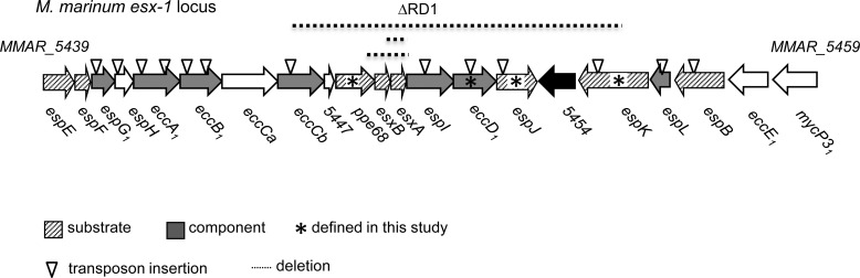
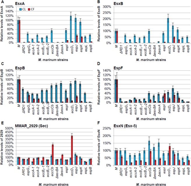
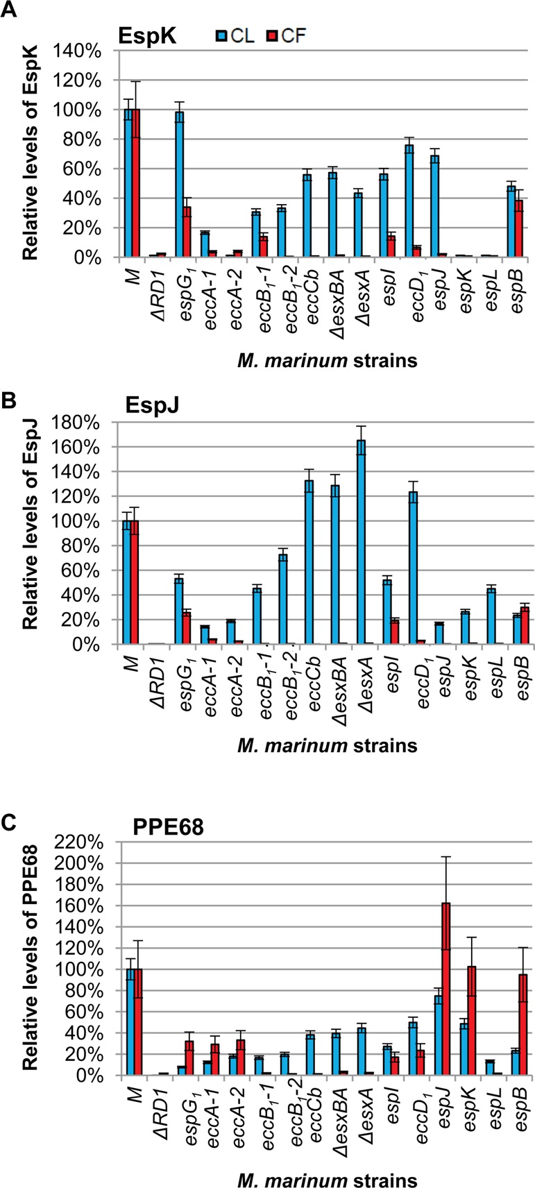
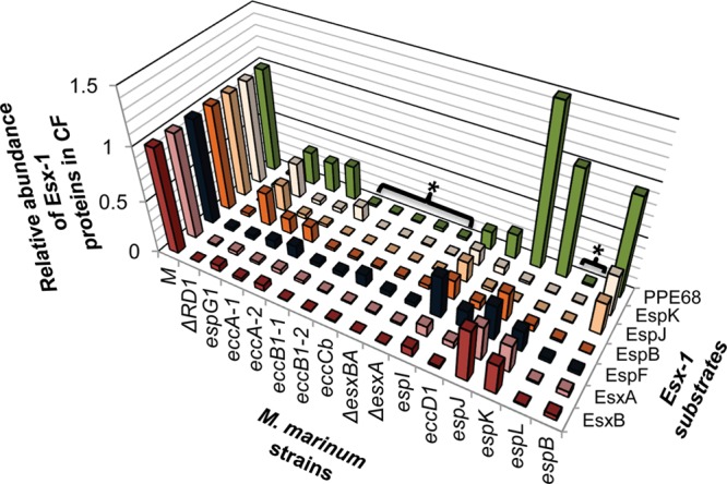
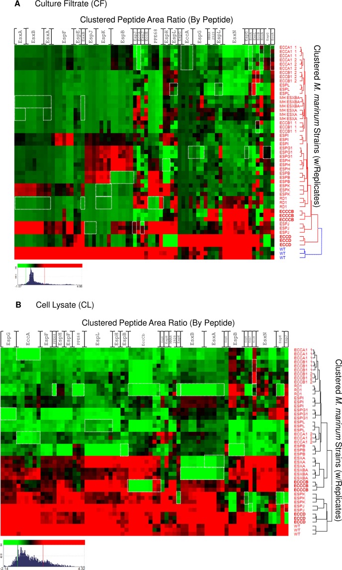
The Esx/WXG-100 (ESAT-6/Wss) exporters are multiprotein complexes that promote protein translocation across the cytoplasmic membrane in a diverse range of pathogenic and nonpathogenic bacterial species. The Esx-1 (ESAT-6 System-1) system mediates virulence factor translocation in mycobacterial pathogens, including the human pathogen Mycobacterium tuberculosis. Although several genes have been associated with Esx-1-mediated transport and virulence, the contribution of individual Esx-1 genes to export is largely undefined. A unique aspect of Esx-1 export is that several substrates require each other for export/stability. We exploited substrate "codependency" to identify Esx-1 substrates. We simultaneously quantified changes in the levels of 13 Esx-1 proteins from both secreted and cytosolic protein fractions generated from 16 Esx-1-deficient Mycobacterium marinum strains in a single experiment using MRM/SRM targeted mass spectrometry. This expansion of measurable Esx-1 proteins allowed us to define statistical rules for assigning novel substrates using phenotypic profiles of known Esx-1 substrates. Using this approach, we identified three additional Esx-1 substrates encoded by the esx-1 region. Our studies begin to address how disruption of specific genes affects several proteins in the Esx-1 complex. Overall, our findings illuminate relationships between Esx-1 proteins and create a framework for the identification of secreted substrates applicable to other protein exporters and pathways.

Keywords: Esx-1; EsxA; MRM/SRM; Mycobacterium marinum; RD1; secretion; substrate identification; targeted proteomics.


Figures

Figure 1
Figure 1
The esx-1 region in M. marinum. The ∼10 Kb genomic region including the Esx-1 secretion system from M. marinum is shown. Genes are named according to Bitter et al. Briefly, Esp = Esx secretion-associated proteins; Ecc = Esx conserved components. Subscript “1” indicates the gene is found at the esx-1 locus. Genes include letters based on the order of the genes at the esx-1 locus. Transposon (Tn) insertion strains used in this study are represented with an inverted open triangle. Strains used in this study are in Supplemental Table S1. Deletions are indicated by a dotted line above the genes within the deletion. MMAR_5454 is not conserved in M. tuberculosis. * indicates substrates or components defined in this study.
Figure 2
Figure 2
Development and validation of the targeted MS Esx-1 secretion assay. (A) nLC–MRM analysis of EsxA. Relative EsxA levels within the cell lysate (CL, blue bars) or in the culture filtrate (CF, red bars) generated from Esx-1-deficient strains as compared to wild-type M. marinum. (B) nLC–MRM analysis of EsxB. For EsxA and EsxB, the ΔRD1, ΔesxA, and ΔesxBA strains served as negative controls for EsxA and EsxB detection and demonstrated the specificity of the approach. (C) nLC–MRM analysis of the levels of the EspB substrate in the CL and CF relative to the levels of EspB in the wild-type strain. For EspB, the espB::Tn strain served as a negative control for EspB detection and demonstrated the specificity of the approach. (D) nLC–MRM analysis of the levels of the EspF substrate in the CL and CF. (E) nLC–MRM analysis of MMAR_2929. (F) nLC–MRM analysis of EsxN. Error bars represent the average propagated standard error and were calculated as described in the Experimental Procedures. In all graphs changes in the levels of protein in each of the Esx-1-deficient strains as compared to the wild-type strain were considered significantly different from each other if the p value from a two-tailed Student’s t test was ≤0.05. Changes in protein levels between the wild-type and each Esx-1-deficient strain that were not statistically significant (p ≥ 0.05) were as follows: (A) The levels of EsxA in the cell lysates from the espI, eccD1, espJ, and espK strains were not statistically different from the levels in the wild-type strain. (B) The levels of EsxB in the cell lysates from the eccD1, espJ, and espK strains were not statistically different from the levels in the wild-type strain. (C) The levels of EspB in the cell lysates from the eccB1-2, eccCb, esxBA, esxA, eccD1, espK, and espL were not significantly different from the levels in the wild-type strain. (F) The levels of EsxN in the lysates from the ΔRD1, espG1, eccB1-2, espI, eccD1, espJ, espK, espL, and espB strains were not statistically different from the wild-type strain. The levels of EsxN in the culture filtrate generated from the eccCb, eccD1, and espJ strains were not statistically different from the WT strain. The actual p values are listed in the Supporting Information (Table S2). Log2 transformed versions of the CF data are available in Supplemental Figure S4.
Figure 3
Figure 3
EspK, EspJ, and PPE68 are Esx-1 substrates. (A) nLC–MRM analysis of the levels of EspK in cell lysate (CL, blue bars) and culture filtrate (CF, red bars) relative to the levels of EspK in the WT M strain. The ΔRD1 and espK::Tn strains served as a negative control for EspK detection and demonstrate specificity of the approach. (B) nLC–MRM analysis of the levels of the EspJ substrate in the CL and CF. The ΔRD1 strain and espJ::Tn strain serve as a negative control for EspJ detection. We report a weak false positive signal indicating extremely low levels of EspJ peptides in the espJ::Tn strain. One EspJ peptide was observed at <0.5% of the levels in the espB::Tn strain and <0.2% of the EspJ levels observed in the WT M strain. This low false-positive signal was due to the large EspJ signal observed in the espB::Tn strain, which was the previous nLC injection on the mass spectrometer. For example, the EspJ tryptic peptide TSSMSTAADIYAK was present at ∼1e4cps2 in the M strain and was not detected in ΔRD1 or the espJ::Tn strains. AEPLAVDPAR is a high-intensity proteotypic peptide for EspJ that was measured at >2e6cps2 in the WT strain and 1.3e6cps2 in the espB::Tn strain, which was injected immediately prior to espJ::Tn analysis. Further evidence of this is the gradual reduction in carryover signal with each successive analysis of the espJ::Tn strain. (C) nLC–MRM analysis of the levels of PPE68 in the CL and CF. The ΔRD1 strain served as a negative control for PPE68 detection. Error bars represent the average propagated standard error and were calculated as described in the Experimental Procedures. The differences between the levels of each protein in the WT and each Esx-1-deficient strain were considered statistically significant if p values were less than 0.05 based on a two-tailed Student’s t test. The levels of the indicated protein in each Esx-1-deficient strain were significantly different from the wild-type levels with the following exceptions: The levels of EspK in the cell lysates of the espG strain were not different from the levels of EspK in the wild-type lysate. The levels of PPE68 in the cell lysate from the espJ strain were not significantly different from the wild-type strain. The levels of PPE68 in the culture filtrate generated from the espK strain were not significantly different from the levels of PPE68 in the culture filtrate from the wild-type strain. The actual p values are listed in Supplemental Table S2. Log2 transformed versions of the CF data are available in Supplemental Figure S4.
Figure 4
Figure 4
Divergent genetic requirements for substrate export. The graph summarizes the findings presented in Figures 2 and 3. The levels of secreted proteins present in the CF of the Esx-1-deficient strains relative to the WT strain illustrate divergent genetic requirements for substrate export. Brackets with asterisks highlight strains deficient for export of all of the substrates tested, similar to the ΔRD1 strain.
Figure 5
Figure 5
Hierarchical clustering of the Esx-1-associated proteome by peptide. Hierarchical clustering was performed on the quantitative peak area response of Esx-1-derived peptides vs WT and the Esx-1-deficient M. marinum strain library. Red intensity indicates higher expression, and green indicates lower expression vs the mean value for each gene product. Clustering was independently performed on nLC–MRM data from (A) culture filtrate (CF) and (B) cell lysate (CL) peak response. Threshold was set to 1S.D for color response. Displayed dynamic range for CF (Log2 −1.87–4.96 green–red) and CL (Log2 −2.14–4.32 green–red). Novel ESX-1 substrates PPE68, EspJ, and EspK are readily apparent from examination of the CF data, and loss of substrate stability in Esx-1 mutants is also well represented in CL fractions. White boxes indicate peptides that are absent due to interruption or loss of the gene that encodes the protein. For example, in the esxA deletion strains, the peptides corresponding to EsxA are boxed in white. Columns are listed for each protein for simplicity but represent individual peptide responses as in Supplemental Figure S1.

References

    1. BCG Vaccine; World Health Organization: Geneva, 2009.
    1. Tuberculosis & Diabetes: Collaborative framework for care and control of tuberculosis and diabetes; World Health Organization: Geneva, 2011. - PubMed
    1. WHO Global Tuberculosis Report 2013; World Health Organization: Geneva, 2013.
    1. Burts M. L.; Williams W. A.; DeBord K.; Missiakas D. M. EsxA and EsxB are secreted by an ESAT-6-like system that is required for the pathogenesis of Staphylococcus aureus infections. Proc. Natl. Acad. Sci. U.S.A. 2005, 10241169–74. - PMC - PubMed
    1. Garufi G.; Butler E.; Missiakas D. ESAT-6-like protein secretion in Bacillus anthracis. J. Bacteriol. 2008, 190217004–11. - PMC - PubMed

MeSH terms

Substances