. 2015 Mar-Apr;29(3):268-77.
doi: 10.1177/1545968314543500. Epub 2014 Aug 8.

Hindlimb stretching alters locomotor function after spinal cord injury in the adult rat

Affiliations

Hindlimb stretching alters locomotor function after spinal cord injury in the adult rat

Krista L Caudle et al. Neurorehabil Neural Repair. 2015 Mar-Apr.

Abstract

Background: Stretching is a widely accepted standard-of-care therapy following spinal cord injury (SCI) that has not been systematically studied in animal models.

Objective: To investigate the influence of a daily stretch-based physical therapy program on locomotor recovery in adult rats with moderate T9 contusive SCI.

Methods: A randomized treatment and control study of stretching in an animal model of acute SCI. Moderate SCIs were delivered with the NYU Impactor. Daily stretching (30 min/day, 5 days/wk for 8 weeks) was provided by a team of animal handlers. Hindlimb function was assessed using the BBB Open Field Locomotor Scale and kinematically. Passive range-of-motion for each joint was determined weekly using a goniometer.

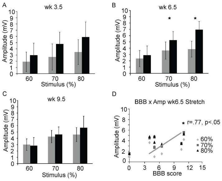
Results: Declines in hindlimb function during overground stepping were observed for the first 4 weeks for stretched animals. BBB scores improved weeks 5 to 10 but remained below the control group. Stretched animals had significant deficits in knee passive range of motion starting at week 4 and for the duration of the study. Kinematic assessment showed decreased joint excursion during stepping that partially recovered beginning at week 5.

Conclusion: Stretch-based therapy significantly impaired functional recovery in adult rats with a moderate contusive SCI at T10. The negative impact on function was greatest acutely but persisted even after the stretching ceased at 8 weeks postinjury.

Keywords: hindlimb stretching; locomotor recovery; physical therapy; spinal cord injury.


Figures

Figure 1
Figure 1. Kinematic Representation of Hindlimb Stretch Positions
A–F, Static passive hindlimb stretch was achieved by manual manipulation of the hindlimb joints. Shown is a three-dimensional (two camera views) kinematic representation of the right hindlimb of each stretch position. The anatomical landmarks represented are the iliac crest (IC), hip, knee, ankle and toe. In order to stretch the ankle flexors (tibialis anterior or TA) the ankle was positioned in extension (A). For the bifunctional muscles rectus femoris (assists with both hip flexion and knee extension), biceps femoris (assists with both hip extension and knee flexion), and the gastrocnemius (assists with both knee flexion and ankle extension), stretch maneuvers required specific positioning of 2 joints as follows: For the triceps surae (lateral and medial gastrocnemius and the soleus), the ankle was positioned in flexion in combination with knee extension, and care was taken to avoid the Achilles tendon with the index finger (B). For the posterior biceps femoris and semitendinosis) the knee was put in extension in combination with hip flexion. For the vastus lateralis, intermedius, medialis and rectus femoris the hip was put in extension in combination with knee flexion (C,D). In order to stretch the hip adductor muscles (adductors brevis, longus, magnus, minimus, pectineus, gracilis and obturator) hip abduction was achieved by pressing both knees laterally at the same time. Oppositely, the hip abductors (gluteus, iliopsoas) were stretched by crossing one hindlimb over the other medially via stabilizing the hips and placing pressure on the thighs/knees (E,F).
Figure 2
Figure 2. BBB & LSS Scores Daily Stretch
In all graphs (A–D) the bold red x-axis indicates when the stretching protocol was being applied to the acute stretch (SR; A, B & D) and chronic stretch (SR; C) groups. BBB testing was performed three times weekly for all experimental groups: Monday morning (am), Monday afternoon and Friday afternoon , shown in A. Only Monday and Friday pm scores are shown in B. All animals scored 21 pre-injury. Significantly lower BBB scores were found for the SR group (n=7) as compared to non-SR controls during the 8 weeks of daily hindlimb stretch (*) as well as at chronic time points beyond 10 weeks. Animals receiving the daily SR protocol showed dramatic but transient loss of locomotor function until ~5 weeks after which BBB scores gradually increased (weeks 5–8) and reached a plateau (BBB= 11) at 9 weeks after daily stretch ended. C shows the chronic SR group (a sub-group of the controls, n=6) that received the stretch protocol daily for 6 days at 10 weeks (grey trace). Significant loss of function for the chronic SR group occurred after only two consecutive days of stretching, as well as immediately following SR on the following Monday morning. In D, 8 weeks of daily SR protocol, reduced the LSS scores for the SR group indicating a deficit in swimming ability as compared to non-SR controls over weeks 2 and 4. The SR group experienced a gain of swimming function over weeks 6–10.
Figure 3
Figure 3
A–C. CPI, PSI and RI measures for SR animals during shallow water walking at week 3 indicating that they were dragging their hindlimbs, with no swing or stance phases due to lack of weight support. A and C. By 13 weeks, the CPI and RI indices of gait had recovered for SR animals. C. However, the RI shows that significant deficits still exist if the correct pattern is dependent upon plantar steps only. B. Importantly, the ratio of plantar stepping by the hindlimbs (compared to the forelimbs) was delayed but recovered by 13 weeks, indicating that the SR animals were able to weight support and achieved proper plantar paw placement in a few weeks without the daily stretch protocol.
Figure 4
Figure 4. Magnetically Evoked Muscle Responses Daily Stretch
A–C. Bilateral EMG recordings from lateral gastrocnemius were assessed after BBB scoring on Monday afternoon at weeks 3.5, 6.5 and 9.5. B. Week 6.5 showed significantly lower amplitude for SR group as compared to non-SR controls. D. There was a positive correlation for evoked response amplitude and BBB scores at week 6.5

References

    1. O’sullivan, Schmitz Guide to Physical Therapist Practice, 2nd Edition. Phys Ther. 2001;81:9–746. - PubMed
    1. Harvey LA, Glinsky JA, Katalinic OM, Ben M. Contracture management for people with spinal cord injuries. NeuroRehabilitation. 2011;28(1):17–20. - PubMed
    1. Schieppati M, Romano C, Gritti I. Convergence of Ia fibres from synergistic and antagonistic muscles onto interneurones inhibitory to soleus in humans. J Physiol. 1990;431:365–377. - PMC - PubMed
    1. Harvey LA, Herbert RD, Glinsky J, Moseley AM, Bowden J. Effects of 6 months of regular passive movements on ankle joint mobility in people with spinal cord injury: a randomized controlled trial. Spinal Cord. 2009;47(1):62–66. - PubMed
    1. Harvey LA, Herbert RD. Muscle stretching for treatment and prevention of contracture in people with spinal cord injury. Spinal Cord. 2002;40(1):1–9. - PubMed

Publication types