. 2014 Dec 1;59(11):1638-47.
doi: 10.1093/cid/ciu641. Epub 2014 Aug 8.

Neutrophil-associated central nervous system inflammation in tuberculous meningitis immune reconstitution inflammatory syndrome

Affiliations

Neutrophil-associated central nervous system inflammation in tuberculous meningitis immune reconstitution inflammatory syndrome

Suzaan Marais et al. Clin Infect Dis. .

Abstract

Background: The immunopathogenesis of tuberculosis-associated immune reconstitution inflammatory syndrome (IRIS) remains incompletely understood, and we know of only 1 disease site-specific study of the underlying immunology; we recently showed that Mycobacterium tuberculosis culture positivity and increased neutrophils in the cerebrospinal fluid (CSF) of patients with tuberculous meningitis (TBM) are associated with TBM-IRIS. In this study we investigated inflammatory mediators at the disease site in patients with TBM-IRIS.

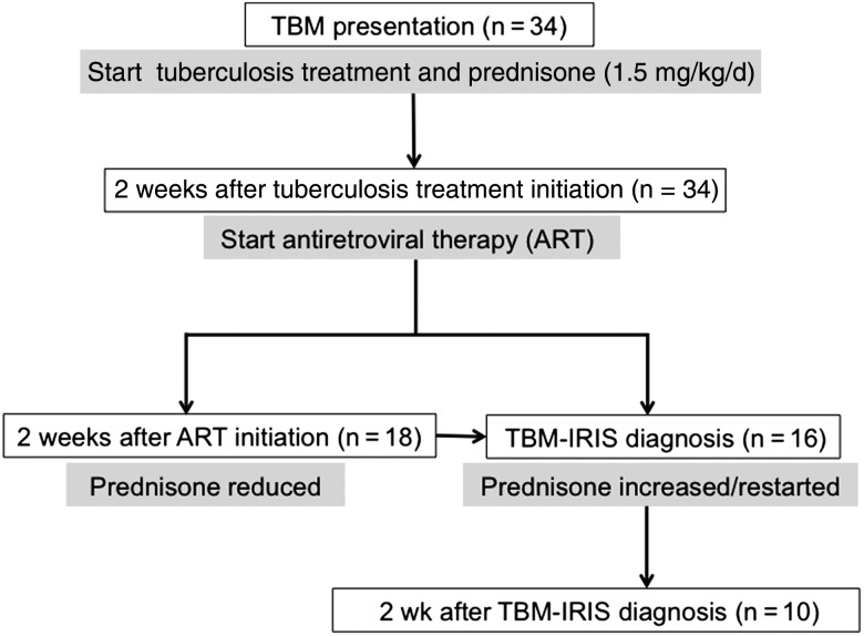
Methods: We performed lumbar puncture at 3-5 time points in human immunodeficiency virus (HIV)-infected patients with TBM (n = 34), including at TBM diagnosis, at initiation of antiretroviral therapy (ART) (day 14), 14 days after ART initiation, at presentation of TBM-IRIS, and 14 days thereafter. We determined the concentrations of 40 mediators in CSF (33 paired with blood) with Luminex or enzyme-linked immunosorbent assays. Findings were compared between patients who developed TBM-IRIS (n = 16) and those who did not (TBM-non-IRIS; n = 18).

Results: At TBM diagnosis and 2 weeks after ART initiation, TBM-IRIS was associated with severe, compartmentalized inflammation in the CSF, with elevated concentrations of cytokines, chemokines, neutrophil-associated mediators, and matrix metalloproteinases, compared with TBM-non-IRIS. Patients with TBM-non-IRIS whose CSF cultures were positive for M. tuberculosis at TBM diagnosis (n = 6) showed inflammatory responses similar to those seen in patients with TBM-IRIS at both time points. However, at 2 weeks after ART initiation, S100A8/A9 was significantly increased in patients with TBM-IRIS, compared with patients with TBM-non-IRIS whose cultures were positive at baseline.

Conclusions: A high baseline M. tuberculosis antigen load drives an inflammatory response that manifests clinically as TBM-IRIS in most, but not all, patients with TBM. Neutrophils and their mediators, especially S100A8/A9, are closely associated with the central nervous system inflammation that characterizes TBM-IRIS.

Keywords: HIV; antiretroviral therapy; therapy-complications; tuberculosis.


Figures

Figure 1.
Figure 1.
Flow diagram of time points when lumbar puncture and phlebotomy were performed in patients with tuberculous meningitis (TBM). Drug interventions are indicated at each time point (gray boxes). Procedures were performed at a minimum of 3 time points: (1) TBM diagnosis, (2) antiretroviral treatment (ART) initiation, and (3) 2 weeks after ART initiation or TBM immune reconstitution inflammatory syndrome (IRIS) presentation, whichever occurred first. Patients in whom TBM-IRIS developed later than 2 weeks after ART initiation underwent repeated procedures at TBM-IRIS presentation. Unless lumbar puncture was contraindicated, procedures were repeated in patients with TBM-IRIS 2 weeks after TBM-IRIS presentation. At the start of the study, the first-line ART regimen for patients receiving tuberculosis treatment in South Africa was stavudine, lamivudine, and efavirenz (started in 10 patients [63%] with TBM-IRIS and 10 [56%] with TBM without IRIS [TBM-non-IRIS]). Later during the study, tenofovir replaced stavudine, according to revised national guidelines (started in 4 patients [25%] with TBM-IRIS and 5 [28%] with TBM-non-IRIS). Five patients (2 [13%] with TBM-IRIS and 3 [17%] with TBM-non-IRIS), in whom these regimens were contraindicated, received zidovudine, lamivudine, and efavirenz.
Figure 2.
Figure 2.
Box plots of mediator concentrations over time in cerebrospinal fluid of patients who developed tuberculous meningitis (TBM) immune reconstitution inflammatory syndrome (IRIS) (red) and those who did not (blue). The assay limits of detection have been substituted for 0 values. The left y-axis is a log10 scale, and the right y-axis indicates time points of sample collection. Within graphs, boxes with horizontal lines represent interquartile ranges (IQR) and medians, and vertical line represent 95% confidence intervals. Data points for outliers (≥1.5 × IQR) are included. (See Supplementary Tables 3–5 for P values of analyses between groups.) For patients with TBM-IRIS, the “2 wk after antiretroviral therapy (ART)” time point indicates findings at TBM-IRIS presentation, which occurred a median of 14 days (interquartile range, 4–20 days) after initiation of ART. Concentrations of all mediators were measured as picograms per milliliter, with the exception of cathepsin G, which was measured in units per milliliter. Abbreviations: C5a, complement 5a; CCL, CC chemokine ligand; CXCL, CXC chemokine ligand; G-CSF, granulocyte colony-stimulating factor; GM-CSF, granulocyte-macrophage colony-stimulating factor; HNP, human neutrophil peptide; IFN, interferon; IL, interleukin; MMP, metalloproteinase; TIMP, tissue inhibitor of MMP; TNF, tumor necrosis factor.
Figure 3.
Figure 3.
Representative examples of changes over time of cerebrospinal fluid (CSF) mediators in patients who developed tuberculous meningitis (TBM) immune reconstitution inflammatory syndrome (IRIS) (n = 16) (red) and those who did not (n = 18) (blue). For patients with TBM-IRIS, the “2 wk after ART [antiretroviral therapy]” time point indicates findings at TBM-IRIS presentation. Some patients with TBM without IRIS (TBM-non-IRIS) showed an increase in mediator concentration and the ratio of metalloproteinase (MMP) 9 to tissue inhibitor of MMP tissue inhibitor of MMP (TIMP) 1 after starting ART; these patients had CSF cultures positive for Mycobacterium tuberculosis at TBM diagnosis. Abbreviations: CXCL, CXC chemokine ligand; HNP, human neutrophil peptide; IL, interleukin; TNF, tumor necrosis factor.
Figure 4.
Figure 4.
Unsupervised hierarchical clustering of tuberculous meningitis (TBM) at TBM diagnosis (A), initiation of antiretroviral therapy (ART) (B), and TBM immune reconstitution inflammatory syndrome (IRIS) presentation or 2 weeks after ART initiation (patients with non-TBM-IRIS) (C). IRIS status indicates patients who developed TBM-IRIS (red) and those who did not (blue). Culture status indicates cerebrospinal fluid cultures positive (black) or negative (green) for Mycobacterium tuberculosis at TBM diagnosis. Values plotted are natural log + 1. Corresponding concentrations in picograms per milliliter are indicated on the color key. Concentrations of all mediators were measured in picograms per milliliter, with the exception of cathepsin G, measured in units per milliliter; neutrophils and lymphocytes were measured as cells × 106 per liter. Abbreviations: C5a, complement 5a; CCL, CC chemokine ligand; CXCL, CXC chemokine ligand; G-CSF, granulocyte colony-stimulating factor; GM-CSF, granulocyte-macrophage colony-stimulating factor; HNP, human neutrophil peptide; IFN, interferon; IL, interleukin; MMP, metalloproteinase; TIMP, tissue inhibitor of MMP; TNF, tumor necrosis factor.
Figure 4.
Figure 4.
Unsupervised hierarchical clustering of tuberculous meningitis (TBM) at TBM diagnosis (A), initiation of antiretroviral therapy (ART) (B), and TBM immune reconstitution inflammatory syndrome (IRIS) presentation or 2 weeks after ART initiation (patients with non-TBM-IRIS) (C). IRIS status indicates patients who developed TBM-IRIS (red) and those who did not (blue). Culture status indicates cerebrospinal fluid cultures positive (black) or negative (green) for Mycobacterium tuberculosis at TBM diagnosis. Values plotted are natural log + 1. Corresponding concentrations in picograms per milliliter are indicated on the color key. Concentrations of all mediators were measured in picograms per milliliter, with the exception of cathepsin G, measured in units per milliliter; neutrophils and lymphocytes were measured as cells × 106 per liter. Abbreviations: C5a, complement 5a; CCL, CC chemokine ligand; CXCL, CXC chemokine ligand; G-CSF, granulocyte colony-stimulating factor; GM-CSF, granulocyte-macrophage colony-stimulating factor; HNP, human neutrophil peptide; IFN, interferon; IL, interleukin; MMP, metalloproteinase; TIMP, tissue inhibitor of MMP; TNF, tumor necrosis factor.

References

    1. Meintjes G, Wilkinson RJ, Morroni C, et al. Randomized placebo-controlled trial of prednisone for paradoxical tuberculosis-associated immune reconstitution inflammatory syndrome. AIDS. 2010;24:2381–90. - PMC - PubMed
    1. Burman W, Weis S, Vernon A, et al. Frequency, severity and duration of immune reconstitution events in HIV-related tuberculosis. Int J Tuberc Lung Dis. 2007;11:1282–9. - PubMed
    1. Marais S, Meintjes G, Pepper DJ, et al. Frequency, severity, and prediction of tuberculous meningitis immune reconstitution inflammatory syndrome. Clin Infect Dis. 2013;56:450–60. - PMC - PubMed
    1. Meintjes G, Lawn SD, Scano F, et al. Tuberculosis-associated immune reconstitution inflammatory syndrome: case definitions for use in resource-limited settings. Lancet Infect Dis. 2008;8:516–23. - PMC - PubMed
    1. Pepper DJ, Marais S, Maartens G, et al. Neurologic manifestations of paradoxical tuberculosis-associated immune reconstitution inflammatory syndrome: a case series. Clin Infect Dis. 2009;48:e96–107. - PubMed

Publication types

MeSH terms