Review
. 2014 Nov:96:46-54.
doi: 10.1016/j.mvr.2014.07.013. Epub 2014 Aug 5.

Mechanical forces and lymphatic transport

Affiliations
Review

Mechanical forces and lymphatic transport

Jerome W Breslin. Microvasc Res. 2014 Nov.

Abstract

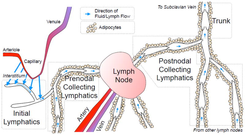
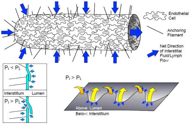
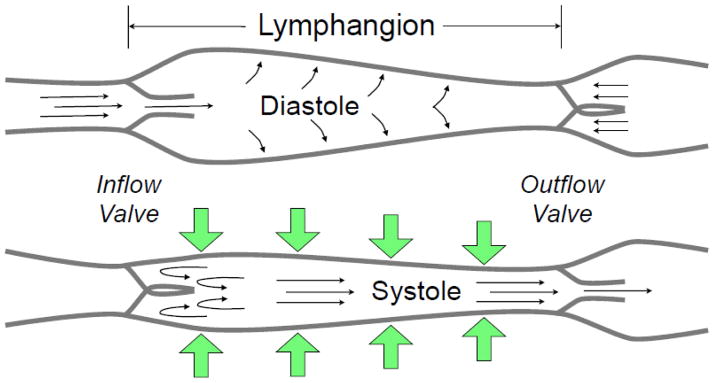
This review examines the current understanding of how the lymphatic vessel network can optimize lymph flow in response to various mechanical forces. Lymphatics are organized as a vascular tree, with blind-ended initial lymphatics, precollectors, prenodal collecting lymphatics, lymph nodes, postnodal collecting lymphatics and the larger trunks (thoracic duct and right lymph duct) that connect to the subclavian veins. The formation of lymph from interstitial fluid depends heavily on oscillating pressure gradients to drive fluid into initial lymphatics. Collecting lymphatics are segmented vessels with unidirectional valves, with each segment, called a lymphangion, possessing an intrinsic pumping mechanism. The lymphangions propel lymph forward against a hydrostatic pressure gradient. Fluid is returned to the central circulation both at lymph nodes and via the larger lymphatic trunks. Several recent developments are discussed, including evidence for the active role of endothelial cells in lymph formation; recent developments on how inflow pressure, outflow pressure, and shear stress affect the pump function of the lymphangion; lymphatic valve gating mechanisms; collecting lymphatic permeability; and current interpretations of the molecular mechanisms within lymphatic endothelial cells and smooth muscle. An improved understanding of the physiological mechanisms by which lymphatic vessels sense mechanical stimuli, integrate the information, and generate the appropriate response is key for determining the pathogenesis of lymphatic insufficiency and developing treatments for lymphedema.

Keywords: Lymphatic contractile cycle; Lymphatic endothelium; Lymphatic muscle; Lymphatic myogenic response; Lymphedema.


Figures

Fig. 1
Fig. 1
Schematic view of lymph formation and transport. The interstitial fluid formed by capillary filtration moves toward initial lymphatics (large blue arrows). This fluid then moves along the initial lymphatic network into contractile prenodal collecting lymphatics (small blue arrows). The collecting lymphatics frequently are surrounded by adipocytes, and bring lymph to one or more lymph nodes, where some of the lymph fluid is lost to the blood circulation. A single postnodal collecting lymphatic exits a lymph node, eventually coalescing with other collecting lymphatics into a larger lymph trunk. The two largest, main trunks of this vascular tree (thoracic duct, right lymph duct) empty into the left and right subclavian veins, respectively.
Fig. 2
Fig. 2
Initial lymphatics and lymph formation. The initial lymphatics are composed of a single layer of oak-leaf shaped endothelial cells with discontinuous basement membrane and anchoring filaments that project into the interstitium, and are the site of interstitial fluid entry (blue arrows). Fluid can pass through leaflets of cell membrane at adjacent endothelial cells when the interstitial pressure (Pi) exceeds luminal pressure (PL). The leaflets are closed when PL > Pi (bottom left; light blue arrows represent net pressure gradient), and open when PL < Pi. The sites where these leaflets are thought to open are the along the far edges of the “oak leaf” junction lobes (bottom right). The yellow arrows show the direction of the leaflet movement when PL < Pi and the small blue arrows show where fluid is thought to pass across these specialized lymphatic endothelial junctions.
Fig. 3
Fig. 3
Collecting lymphatic phasic contractions. Like the heart, collecting lymphatics have a period of brisk contraction (systole) and a period of relaxation (diastole) between each phasic contraction. Each lymphangion, defined as the segment between two unidirectional valves, can typically exhibit systole and diastole. When a lymphangion is relaxed, the inflow (or upstream) valve will open (given sufficient inflow pressure) for filling of the lymphangion to occur. During systole, the phasic contraction pushes the lymph both upstream and downstream, but the inflow valve closes, so that lymph is forced forward through the outflow valve. The exact valve gating and phasic contraction frequency and force depend heavily on the outflow pressure (afterload), inflow pressure (preload), and the flow of lymph (shear stress), as discussed in the text.

References

    1. Adair TH, Guyton AC. Modification of lymph by lymph nodes. II. Effect of increased lymph node venous blood pressure. Am J Physiol. 1983;245:H616–22. - PubMed
    1. Adair TH, et al. Quantitation of changes in lymph protein concentration during lymph node transit. Am J Physiol. 1982;243:H351–9. - PubMed
    1. Aldrich MB, et al. Lymphatic abnormalities in the normal contralateral arms of subjects with breast cancer-related lymphedema as assessed by near-infrared fluorescent imaging. Biomed Opt Express. 2012;3:1256–65. - PMC - PubMed
    1. Armenio S, et al. Spontaneous contractility in the human lymph vessels. Lymphology. 1981;14:173–8. - PubMed
    1. Baluk P, et al. Functionally specialized junctions between endothelial cells of lymphatic vessels. J Exp Med. 2007;204:2349–62. - PMC - PubMed

Publication types