. 2014 Oct;80(20):6413-26.
doi: 10.1128/AEM.01771-14. Epub 2014 Aug 8.

An ArsR/SmtB family member is involved in the regulation by arsenic of the arsenite oxidase operon in Thiomonas arsenitoxydans

Affiliations

An ArsR/SmtB family member is involved in the regulation by arsenic of the arsenite oxidase operon in Thiomonas arsenitoxydans

Danielle Moinier et al. Appl Environ Microbiol. 2014 Oct.

Abstract

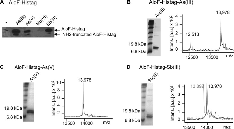
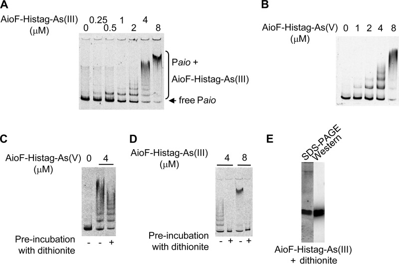
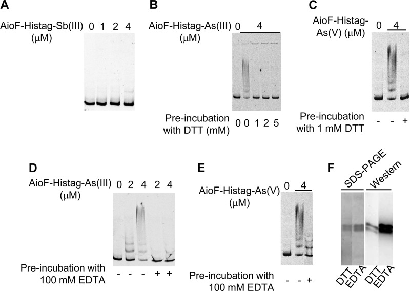
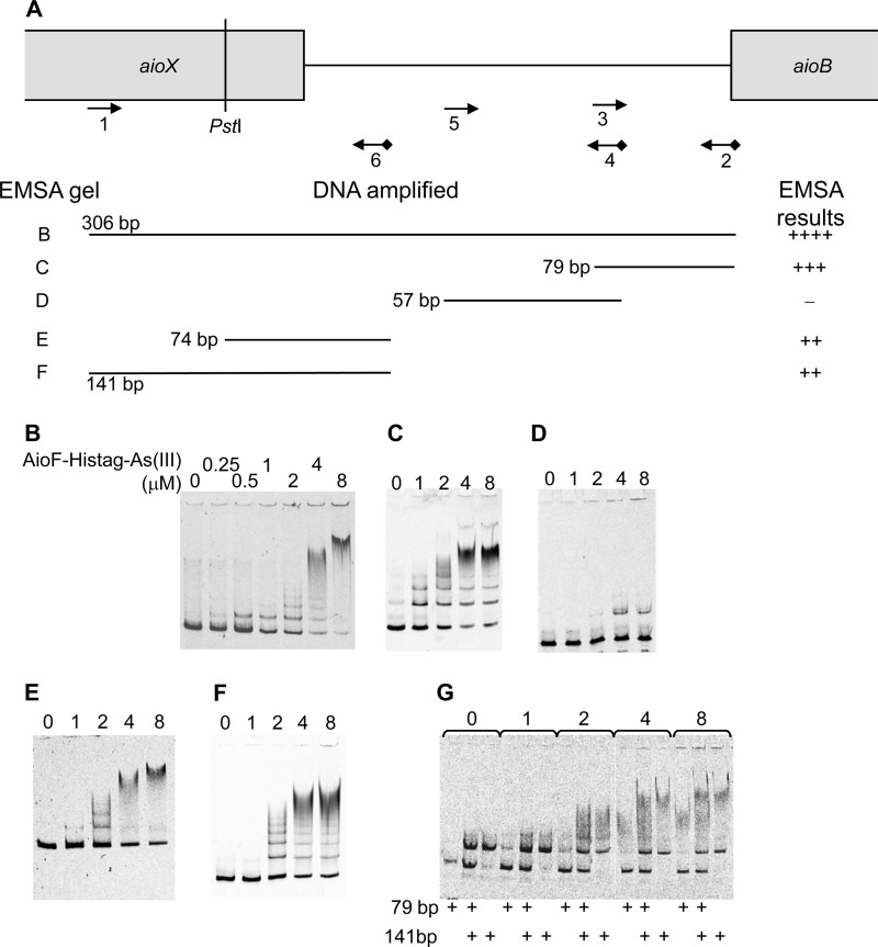
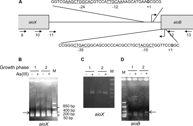
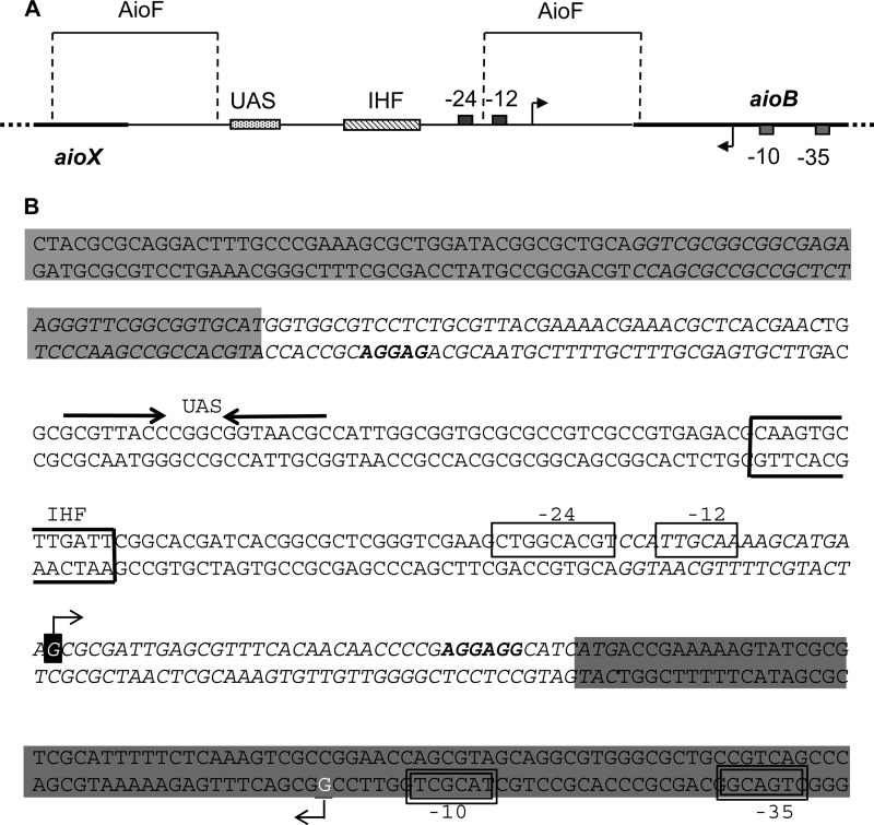
The genetic organization of the aioBA operon, encoding the arsenite oxidase of the moderately acidophilic and facultative chemoautotrophic bacterium Thiomonas arsenitoxydans, is different from that of the aioBA operon in the other arsenite oxidizers, in that it encodes AioF, a metalloprotein belonging to the ArsR/SmtB family. AioF is stabilized by arsenite, arsenate, or antimonite but not molybdate. Arsenic is tightly attached to AioF, likely by cysteine residues. When loaded with arsenite or arsenate, AioF is able to bind specifically to the regulatory region of the aio operon at two distinct positions. In Thiomonas arsenitoxydans, the promoters of aioX and aioB are convergent, suggesting that transcriptional interference occurs. These results indicate that the regulation of the aioBA operon is more complex in Thiomonas arsenitoxydans than in the other aioBA containing arsenite oxidizers and that the arsenic binding protein AioF is involved in this regulation. On the basis of these data, a model to explain the tight control of aioBA expression by arsenic in Thiomonas arsenitoxydans is proposed.


Figures

FIG 1
FIG 1
Analysis of recombinant AioF-His tag produced in Escherichia coli. (A) Total cell extracts obtained in the presence or in the absence of As(III), As(V), Mo(VI), and Sb(III) added at the time of induction with IPTG. AioF-His tag was revealed by immunodetection with antibodies raised against the hexahistidine tag. (B to D) Purified AioF-His tag obtained after addition of As(III) (B), As(V) (C), and Sb(III) (D) at the time of induction with IPTG was analyzed by SDS-PAGE (left) with Coomassie blue staining and MALDI-TOF mass spectrometry (right). Black spectra, reference AioF-His tag-As(III) spectrum (m/z 13,978); gray spectra, reference AioF-His tag-As(V) spectrum (m/z 13,978) in panel C and AioF-His tag-Sb(III) spectrum (m/z 13,892) in panel D. Intens., intensity; a.u., arbitrary units.
FIG 2
FIG 2
Binding of AioF-His tag loaded with As to the aioX-aioB intergenic region analyzed by gel mobility shift assays. A 306-bp DNA fragment encompassing the aioX-aioB intergenic region (A to D) was incubated in the presence of recombinant AioF-His tag-As(III) (A and D) or AioF-His tag-As(V) (B and C). Recombinant AioF-His tag-As(V) (C) and AioF-His tag-As(III) (D) proteins were preincubated with dithionite. (E) SDS-PAGE and Western immunodetection with antibodies against the hexahistidine tag of AioF-His tag-As(III) after dithionite treatment.
FIG 3
FIG 3
Binding of AioF-His tag free of arsenic to the aioX-aioB intergenic region analyzed by gel mobility shift assays. A 306-bp DNA fragment encompassing the aioX-aioB intergenic region was incubated in the presence of the recombinant AioF-His tag-Sb(III) (A), AioF-His tag-As(III) treated with DTT (B) or EDTA (D), or AioF-His tag-As(V) treated with DTT (C) or EDTA (E). (F) SDS-PAGE and Western immunodetection with antibodies against the hexahistidine tag of AioF-His tag-As(III) after DTT or EDTA treatment.
FIG 4
FIG 4
Analysis of AioF-His tag-As(III) binding to different regions of the aioX-aioB intergenic region by gel mobility shift assays. (A) Schematic representation of the DNA fragments analyzed. DNA substrates for band shift assays were produced by PCR amplification using a 5′ Cy5-labeled reverse oligonucleotide (Eurogentec) (see Table S1 in the supplemental material), except that the 74-bp fragment was generated by PstI digestion of the 141-bp amplicon. The locations of the oligonucleotides used to amplify the different DNA fragments are indicated as arrows. The cyanide 5 is indicated as a black diamond. The size of the amplicon, as well as the EMSA gel reference and the EMSA results, is given. The sizes of the different DNA fragments incubated with AioF-His tag-As(III) (0, 0.25, 0.5, 1, 2, 4, or 8 μM) and tested by EMSAs were as follows: 306 bp (B), 79 bp (C), 57 bp (D), 74 bp (E), 141 bp (F), and both 79 and 141 bp (G).
FIG 5
FIG 5
Transcriptional start sites of aioB and aioX operons determined by 5′ RACE. (A) Schematic representation of the aioX-aioB intergenic region. The oligonucleotides used are indicated as arrows. Transcriptional start sites are in bold, and the corresponding σ-dependent promoter motifs are underlined. (B to D) Amplification of cDNA corresponding to the 5′ end of aioX determined in T. arsenitoxydans grown in the presence (lanes +) or the absence (lanes −) of As(III) during the first (lanes labeled 1) and the second (lanes labeled 2) growth phases with oligonucleotide aoxX5′-R2 (arrow labeled 10 in panel A) (B) or oligonucleotide aoxA11 (arrow labeled 11 in panel A) (C). (D) Amplification of cDNA corresponding to the 5′ end of aioB mRNA determined under the same conditions with oligonucleotide aoxA div9 (arrow labeled 12 in panel A). Lanes M, FastRuler low-range DNA ladder (Fermentas).
FIG 6
FIG 6
Predicted structural AioF protein homologs in prokaryotic genomes. (A) Structural and functional model of the AioF protein with Cys residues and the CXXXC motif located upstream and downstream of the HTH region, respectively. (B) Distribution of aioF gene homologs within prokaryotic taxonomic groups. For each main archaeal and bacterial phylum, the numbers in parentheses indicate the number of organisms for which complete genomes were available for this study. G and H, the number of genomes harboring at least one aioF and the total number aioF genes in the phylum, respectively.
FIG 7
FIG 7
Schematic representation (A) and genetic organization (B) of the aioX-aioB intergenic region. aioX and aioB are shaded in light and dark gray, respectively. The ribosome binding sites are in bold letters. The −24 and −12 boxes of the σ54-dependent promoter of aioB are boxed, and the upstream activating sequence (UAS) likely corresponding to the AioR binding site is indicated above the sequence with converging arrows. The −35 and −10 boxes of the σ70-dependent promoter of aioX are boxed with double lines. Transcriptional start sites are indicated in reverse white lettering. The IHF binding site is boxed in bold. The regions where AioF binds are indicated in italics.

References

    1. Bird LJ, Bonnefoy V, Newman DK. 2011. Bioenergetic challenges of microbial iron metabolisms. Trends Microbiol. 19:330–340. 10.1016/j.tim.2011.05.001 - DOI - PubMed
    1. Slyemi D, Bonnefoy V. 2012. How prokaryotes deal with arsenic. Environ. Microbiol. Rep. 4:571–586. 10.1111/j.1758-2229.2011.00300.x - DOI - PubMed
    1. Finney LA, O'Halloran TV. 2003. Transition metal speciation in the cell: insights from the chemistry of metal ion receptors. Science 300:931–936. 10.1126/science.1085049 - DOI - PubMed
    1. Pennella MA, Giedroc DP. 2005. Structural determinants of metal selectivity in prokaryotic metal-responsive transcriptional regulators. Biometals 18:413–428. 10.1007/s10534-005-3716-8 - DOI - PubMed
    1. Osman D, Cavet JS. 2010. Bacterial metal-sensing proteins exemplified by ArsR-SmtB family repressors. Nat. Prod. Rep. 27:668–680. 10.1039/b906682a - DOI - PubMed

Publication types

MeSH terms

LinkOut - more resources