

Glycobiology of ocular angiogenesis
- PMID: 25108228
- PMCID: PMC4211602
- DOI: 10.1093/glycob/cwu078
Glycobiology of ocular angiogenesis
Abstract
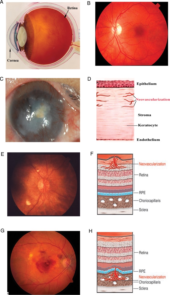
Ocular neovascularization can affect almost all the tissues of the eye: the cornea, the iris, the retina, and the choroid. Pathological neovascularization is the underlying cause of vision loss in common ocular conditions such as diabetic retinopathy, retinopathy of prematurity and age-related macular neovascularization. Glycosylation is the most common covalent posttranslational modification of proteins in mammalian cells. A growing body of evidence demonstrates that glycosylation influences the process of angiogenesis and impacts activation, proliferation, and migration of endothelial cells as well as the interaction of angiogenic endothelial cells with other cell types necessary to form blood vessels. Recent studies have provided evidence that members of the galectin class of β-galactoside-binding proteins modulate angiogenesis by novel carbohydrate-based recognition systems involving interactions between glycans of angiogenic cell surface receptors and galectins. This review discusses the significance of glycosylation and the role of galectins in the pathogenesis of ocular neovascularization.
Keywords: angiogenesis; choroidal neovascularization; corneal neovascularization; galectin-3; glycans; glycosylation; integrins; neovascularization; retinal neovascularization.
© The Author 2014. Published by Oxford University Press. All rights reserved. For permissions, please e-mail: journals.permissions@oup.com.
Figures
References
-
- Adamis AP, Aiello LP, D'Amato RA. Angiogenesis and ophthalmic disease. Angiogenesis. 1999;3:9–14. - PubMed
-
- Adamis AP, Miller JW, Bernal MT, D'Amico DJ, Folkman J, Yeo TK, Yeo KT. Increased vascular endothelial growth factor levels in the vitreous of eyes with proliferative diabetic retinopathy. Am J Ophthalmol. 1994;118:445–450. - PubMed
-
- Afzal A, Shaw LC, Ljubimov AV, Boulton ME, Segal MS, Grant MB. Retinal and choroidal microangiopathies: Therapeutic opportunities. Microvasc Res. 2007;74:131–144. - PubMed
-
- Aiello LP, Avery RL, Arrigg PG, Keyt BA, Jampel HD, Shah ST, Pasquale LR, Thieme H, Iwamoto MA, Park JE, et al. Vascular endothelial growth factor in ocular fluid of patients with diabetic retinopathy and other retinal disorders. N Engl J Med. 1994;331:1480–1487. - PubMed
-
- Asahara T, Chen D, Takahashi T, Fujikawa K, Kearney M, Magner M, Yancopoulos GD, Isner JM. Tie2 receptor ligands, angiopoietin-1 and angiopoietin-2, modulate VEGF-induced postnatal neovascularization. Circ Res. 1998;83:233–240. - PubMed
Publication types
MeSH terms
Substances
Grants and funding
LinkOut - more resources
Full Text Sources
Other Literature Sources
