. 2014 Sep;20(9):1018-26.
doi: 10.1038/nm.3587. Epub 2014 Aug 10.

An acetate switch regulates stress erythropoiesis

Affiliations

An acetate switch regulates stress erythropoiesis

Min Xu et al. Nat Med. 2014 Sep.

Abstract

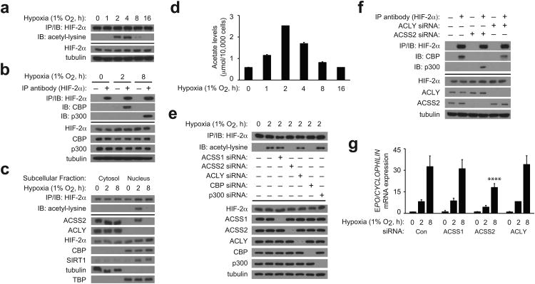
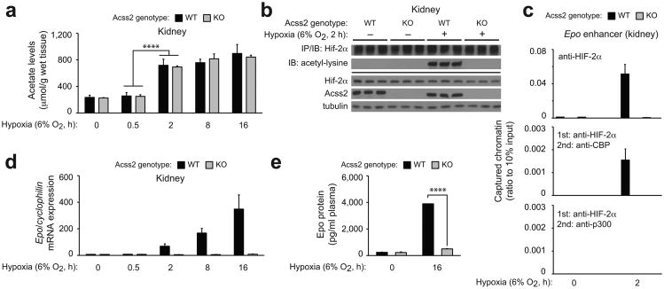
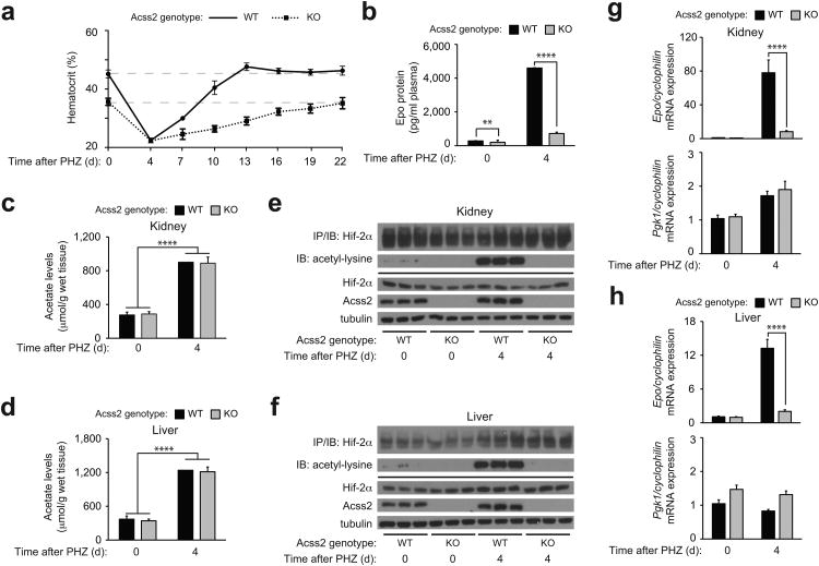
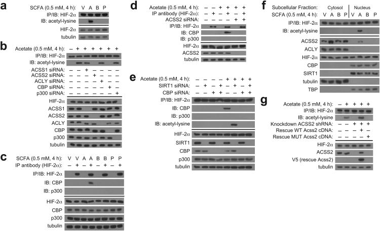
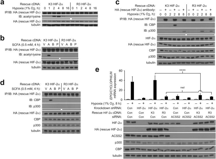
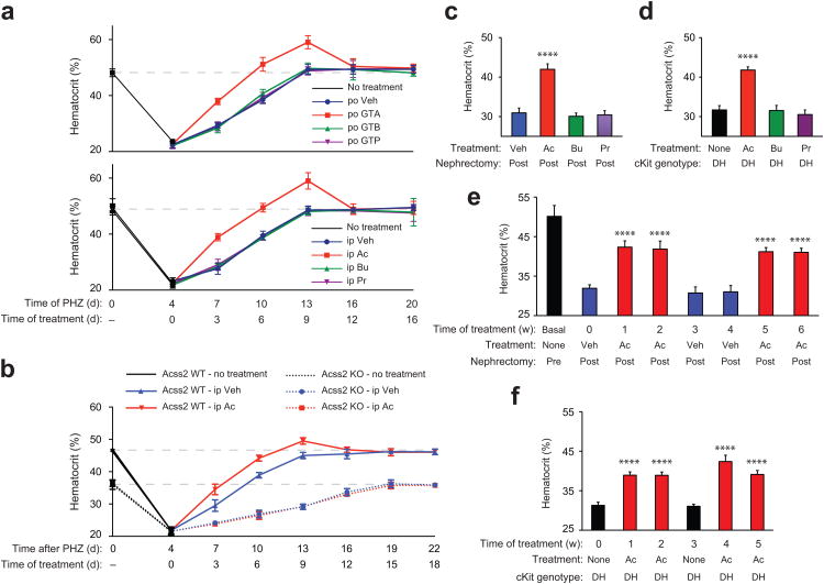
The hormone erythropoietin (EPO), which is synthesized in the kidney or liver of adult mammals, controls erythrocyte production and is regulated by the stress-responsive transcription factor hypoxia-inducible factor-2 (HIF-2). We previously reported that the lysine acetyltransferase CREB-binding protein (CBP) is required for HIF-2α acetylation and efficient HIF-2-dependent EPO induction during hypoxia. We now show that these processes require acetate-dependent acetyl CoA synthetase 2 (ACSS2). In human Hep3B hepatoma cells and in EPO-generating organs of hypoxic or acutely anemic mice, acetate levels rise and ACSS2 is required for HIF-2α acetylation, CBP-HIF-2α complex formation, CBP-HIF-2α recruitment to the EPO enhancer and efficient induction of EPO gene expression. In acutely anemic mice, acetate supplementation augments stress erythropoiesis in an ACSS2-dependent manner. Moreover, in acquired and inherited chronic anemia mouse models, acetate supplementation increases EPO expression and the resting hematocrit. Thus, a mammalian stress-responsive acetate switch controls HIF-2 signaling and EPO induction during pathophysiological states marked by tissue hypoxia.


Conflict of interest statement

The authors declare no competing financial interests.

Figures

Figure 1
Figure 1. Acss2 controls HIF-2 signaling in hypoxic cells
(a) Time course of endogenous HIF-2α acetylation during hypoxia following immunoprecipitation (IP) of HIF-2α from whole cell extracts and detection of acetylated lysines by immunoblotting (IB). (b) Endogenous CBP/HIF-2α or p300/HIF-2α complex formation after hypoxia exposure. (c) Subcellular localization of ACSS2, ACLY, HIF-2α, CBP, and SIRT1 following hypoxia exposure. Also shown is endogenous acetylated HIF-2α present in each fraction. (d) Acetate levels in cells during hypoxia (n=3 biological replicates/time-point; single measurement/replicate; mean/SD). (e) Contribution of ACSS1, ACSS2, ACLY, CBP and p300 to endogenous HIF-2α acetylation during early (2 h) hypoxia. (f) Contribution of ACLY and ACSS2 to endogenous CBP/HIF-2α or p300/HIF-2α complex formation during early (2 h) hypoxia. (g) Contribution of ACSS1, ACSS2, and ACLY to EPO gene expression following hypoxia exposure (single pool of triplicate biological replicates/manipulation; triplicate measurements/pool; mean/SD). Separate analyses of 0, 2, and 8 h hypoxia samples was performed by two-way ANOVA with Dunnett's multiple comparison post-hoc test using control siRNA as reference within each group; only results for 8 h hypoxia are indicated (****P≤0.0001). All experiments were performed with Hep3B cells and key aspects of the above findings have been confirmed by at least one additional independent experiment.
Figure 2
Figure 2. Acss2 regulates hypoxia-induced renal Epo expression in mice
(a) Acetate levels in kidneys of Acss2 wild-type (WT) or knockout (KO) mice exposed to normoxia or hypoxia (n=3 biological replicates/group, 3 male mice for 8 h time-point, 2 male and 1 female mice for other time-points; single measurements/replicate; mean/SD). Comparison of means was performed by two-way ANOVA with Dunnett's multiple comparison post-hoc test (****P≤0.0001). (b) Acetylation of endogenous HIF-2α isolated by immunoprecipitation (IP) from kidney extracts of Acss2 WT or KO mice and detected by immunoblotting (IB) with anti-acetylated lysine or anti-HIF-2α antibodies. (c) Recruitment of HIF-2α as well as coupled recruitment of HIF-2α with CBP or p300 to the Epo enhancer in kidneys of mixed strain Acss2 WT or KO mice maintained under normoxia or short-term (2 h) continuous hypoxia (n=4 biological replicates/group, 2 male and 2 female mice/group; duplicate measurements/replicate; mean/SEM). (d) Epo gene expression in kidneys of Acss2 KO and WT mice following normoxia or hypoxia exposure (n=3 biological replicates/group, 3 male mice for 8 h time-point, 2 male and 1 female mice for other time-points; triplicate measurements/replicate; mean/SEM). (e) Epo protein measurements of plasma from Acss2 KO and WT mice housed under normoxia or 16 h hypoxia (n=3 biological replicates/group; 2 male, 1 female mice/group; duplicate measurements/replicate; mean/SEM). All comparisons by unpaired Student's t-test with Holm-Sidak correction (****P≤0.0001). Key aspects of the above findings have been confirmed by at least one additional independent experiment, except for (c).
Figure 3
Figure 3. Acute anemia induces Acss2-dependent HIF-2 signaling in mice
(a) Serial hematocrits (n=6 biological replicates/group, 4 male and 2 female mice/group; single measurements/replicate; mean/SD) and (b) plasma Epo protein measurements (n=8 biological replicates/group, 8 male mice/group; duplicate measurements/replicate; mean/SEM) of mixed strain Acss2 wild-type (WT) or knockout (KO) mice after phenylhydrazine (PHZ) treatment. Acetate levels in (c) kidneys and (d) livers of Acss2 WT or KO mice following PHZ treatment (n=10 biological replicates/group, 5 male and 5 female mice/group; single measurements/replicate; mean/SD). All comparisons by unpaired Student's t-test with Holm-Sidak correction (**P≤0.01, ****P≤0.0001). Immunoblot (IB) of acetylated lysine residues following immunoprecipitation (IP) with anti-HIF-2α antibody using (e) kidney or (f) liver extracts derived from three Acss2 WT or KO mice chosen at random from each group in (b). Epo and Pgk1 gene expression in (g) kidneys and (h) livers of same mice in (c) and (d) (n=10 biological replicates/group, 5 male and 5 female mice/group; duplicate measurements/replicate; mean/SEM). All comparisons by two-way ANOVA followed by Tukey's multiple comparison post-hoc test (****P≤0.0001). Key aspects of the above findings have been confirmed by at least one additional independent experiment, except for (c) and (d).
Figure 4
Figure 4. An acetate switch regulates Cbp/HIF-2 interactions in cells
(a) Endogenous HIF-2α acetylation following immunoprecipitation (IP) of endogenous HIF-2α and detection by immunoblotting (IB) with anti-acetylated lysine or anti-HIF-2α antibodies after incubation using medium supplemented with vehicle (V; PBS) or the short chain fatty acid (SCFA) sodium acetate (A), sodium butyrate (B), or sodium propionate (P) prepared in PBS. (b) Contribution of ACSS1, ACSS2, ACLY, CBP and p300 to endogenous HIF-2α acetylation for cells incubated using complete medium supplemented with vehicle or acetate. (c) Endogenous CBP/HIF-2α complexes formed after treatment with SCFA-containing medium. (d) Contribution of ACSS2 to endogenous CBP/HIF-2α complex formation induced by acetate treatment. (e) Endogenous CBP/HIF-2α complexes and acetylated HIF-2α induced by acetate treatment following SIRT1, CBP, or combined SIRT1/CBP knockdown. (f) Subcellular localization of ACSS2, ACLY, HIF-2α, CBP, and SIRT1 following treatment with SCFA-containing medium. (g) Endogenous HIF-2α acetylation induced by acetate supplementation in stably transformed knockdown/rescue cells expressing either an shRNA-resistant wild-type (WT) or mutant (MUT) Acss2 rescue cDNA. All experiments were performed with Hep3B cells. Key aspects of the above findings have been confirmed by at least one additional independent experiment.
Figure 5
Figure 5. Acss2 signaling in cells requires intact HIF-2 acetylation
Acetylation of ectopic rescue wild-type acetylase-sensitive (K3) or acetylase-insensitive (R3) human HIF-2α after immunoprecipitation (IP) of rescue HIF-2α, followed by immunoblotting (IB) of rescue HIF-2α or acetyl-lysine after (a) hypoxia exposure or (b) supplementation of medium with PBS vehicle (V), acetate (A), butyrate (B), or propionate (P). Immunoprecipitation of ectopic rescue K3 or R3 human HIF-2α after (c) hypoxia exposure or (d) supplementation with V, A, B, or P. (e) Measurements of EPO gene expression in stably transformed knockdown/rescue cells expressing ectopic rescue K3 or R3 human HIF-2α and transfected with control or Acss2 siRNA, followed by incubation under either normoxic or hypoxic conditions (single pool of triplicate biological replicates/manipulation, triplicate measurements/pool, mean/SD). All comparisons by two-way ANOVA with Tukey's multiple comparison post-hoc test (nsd, no significant difference; ****P≤0.0001). All experiments were performed with Hep3B cells. Key aspects of the above findings have been confirmed by at least one additional independent experiment.
Figure 6
Figure 6. Acetate facilitates recovery from anemia
(a) Hematocrits of CD1 wild-type female mice after phenylhydrazine (PHZ) treatment, followed by once daily per os (po) supplementation with water vehicle (Veh; n=7 biological replicates), glyceryl triacetate (GTA; n=6 biological replicates), glyceryl tributyrate (GTB; n=8 biological replicates), or glyceryl tripropionate (GTP; n=7 biological replicates) (single measurement/replicate; mean/SD), or followed by once daily intra-peritoneal (ip) supplementation with PBS vehicle (Veh), acetate (Ac), butyrate (Bu), or propionate (Pr) (n=5 biological replicates/group; single measurement/replicate; mean/SD). The dotted line indicates the mean hematocrit for control CD1 wild-type female mice (n=33 biological replicates). (b) Serial hematocrits of mixed strain Acss2 wild-type (WT) or knockout (KO) mice after PHZ treatment, followed by once daily ip Veh or Ac injections (n=4 biological replicates/group except for days 16, 19 and 22 where n=3 biological replicates for Acss2 KO mice treated with Ac following death of one mouse; single measurement/replicate; mean/SD). (c) Hematocrits of CD1 wild-type female mice with chronic renal failure (CRF) induced by 5/6 partial nephrectomy and supplemented with three (Monday, M/Wednesday, W/Friday, F) ip injections of Veh (n=11 biological replicates), Ac (n=11 biological replicates), Bu (n=8 biological replicates), or Pr (n=8 biological replicates) (single measurement/replicate; mean/SD). (d) Hematocrits of WBB6 F1 hybrid double heterozygous (DH) cKit mutant mice with no supplements (None) or supplemented with three (M/W/F) ip injections of Ac, Bu, or Pr (n=4 biological replicates/group; single measurements, mean/SD). (e) Serial hematocrits of CD1 wild-type female mice before (Pre) or after (Post) partial 5/6 nephrectomy, followed by thrice-weekly (M/W/F) ip supplements of Veh or Ac (n=8 biological replicates/group; single measurement/replicate; mean/SD). (f) Serial hematocrits of WBB6 F1 hybrid DH cKit mutant mice without or with thrice-weekly (M/W/F) ip Ac supplementation (n=4 biological replicates/group, 2 male and 2 female mice/group; single measurement/replicate; mean/SD). Hematocrits for (c) through (f) were obtained on the Monday following the indicated treatment period. Key aspects of the above findings have been confirmed by at least one additional independent experiment.

References

    1. Bunn HF, Poyton RO. Oxygen sensing and molecular adaptation to hypoxia. Physiological Reviews. 1996;76:839–885. - PubMed
    1. Richalet JP. Oxygen sensors in the organism: examples of regulation under altitude hypoxia in mammals. Comparative Biochemistry & Physiology, Part A Physiology. 1997;118:9–14. - PubMed
    1. Koury MJ. Erythropoietin: the story of hypoxia and a finely regulated hematopoietic hormone. Exp Hematol. 2005;33:1263–1270. - PubMed
    1. Wang GL, Jiang BH, Rue EA, Semenza GL. Hypoxia-inducible factor 1 is a basic-helix-loop-helix-PAS heterodimer regulated by cellular O2 tension. Proc Natl Acad Sci U S A. 1995;92:5510–5514. - PMC - PubMed
    1. Semenza GL. Regulation of oxygen homeostasis by hypoxia-inducible factor 1. Physiology (Bethesda) 2009;24:97–106. - PubMed

Publication types