. 2014 Aug 11:7:57.
doi: 10.1186/s13041-014-0057-y.

Upregulation of human PINK1 gene expression by NFκB signalling

Upregulation of human PINK1 gene expression by NFκB signalling

Xiaoling Duan et al. Mol Brain. .

Abstract

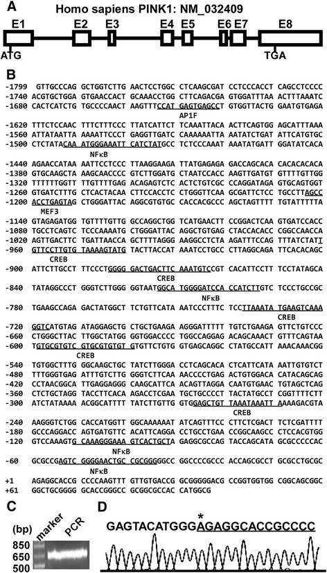
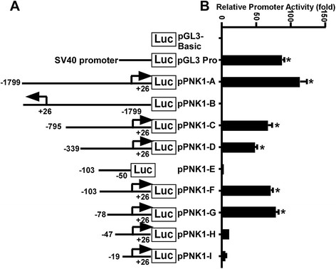
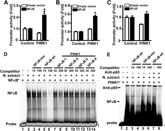
Parkinson's disease (PD) is one of the major neurodegenerative disorders. Mitochondrial malfunction is implicated in PD pathogenesis. Phosphatase and tensin homolog deleted on chromosome 10 (PTEN)-induced putative kinase 1 (PINK1), a serine/threonine kinase, plays an important role in the quality control of mitochondria and more than 70 PINK1 mutations have been identified to cause early-onset PD. However, the regulation of PINK1 gene expression remains elusive. In the present study, we identified the transcription start site (TSS) of the human PINK1 gene using switching mechanism at 5'end of RNA transcription (SMART RACE) assay. The TSS is located at 91 bp upstream of the translation start site ATG. The region with 104 bp was identified as the minimal promoter region by deletion analysis followed by dual luciferase assay. Four functional cis-acting nuclear factor kappa-light-chain-enhancer of activated B cells (NFκB)-binding sites within the PINK1 promoter were identified. NFκB overexpression led to the up-regulation of PINK1 expression in both HEK293 cells and SH-SY5Y cells. Consistently, lipopolysaccharide (LPS), a strong activator of NFκB, significantly increased PINK1 expression in SH-SY5Y cells. Taken together, our results clearly suggested that PINK1 expression is tightly regulated at its transcription level and NFκB is a positive regulator for PINK1 expression.


Figures

Figure 1
Figure 1
Sequence features of the human PINK1 gene promoter. (A) The genomic organization of human PINK1 gene on chromosome 1. E represents exon. ATG is the translation start codon and TGA is the stop codon. (B) The nucleotide sequence of the human PINK1 gene from −1799 to +97 bp. The adenine +1 represents the transcription start site. The putative transcription factor binding sites are underlined in bold face. (C) SMARTer-RACE was performed to map the PINK1 transcription start site. The PCR product was run on a 1.5% agarose gel. (D) The PCR product was cloned into pcDNA4 vector and sequenced to identify the transcription start site. The first base after the adapter is the TSS from which is underlined.
Figure 2
Figure 2
Deletion analysis of the human PINK1 gene promoter. (A) Schematic diagram of the PINK1 promoter constructs consisting of the 5’ flanking region with serial deletions cloned into the pGL3-basic vector. Arrow shows the direction of transcription. The numbers represents the end points of each construct. (B) The deletion plasmids were cotransfected with pCMV-Luc into HEK293 cells. 24 h after the transfection, the luciferase activity was measured and expressed in relative luciferase units (RLU). The pCMV-Luc was used to normalize for transfection efficiency. The values represent means ± SEM. n = 3, *p < 0.0001, by one-way ANOVA followed by post hoc Tukey’s multiple comparisons test.
Figure 3
Figure 3
NFκB binds to human PINK1 gene promoter. The effects of NFκB on the human PINK1 promoter in HEK293 cells (A), SH-SY5Y cells (B) and N2A cells (C) were analyzed by luciferase reporter assays. The PINK1 promoter reporter plasmid (pPINK1-A) or pGL3-promoter was co-transfected into cells with NFκB expression plasmid or the empty vector pMTF. Values indicate means ± SEM. n = 3, *p < 0.01 by Student’s t-test.(D) EMSA with NFκB p65 consensus probe. Lane 1 is labeled probe alone without protein extract. Incubation of the probe with NFκB p65 enriched nuclear extracts forms a shifted DNA-protein complex band (lane 2). Competition assays were performed by further adding different competition oligonucleotides including NFκB-mu, NFκB-wt, and PINK1-NFκB. (E) EMSA with PINK1 NFκB-1 probe. Lane1 is labelled IR700-PINK1-NFκB-1 probe only. Addition of NFκB p65 enriched nuclear extracts forms a shifted DNA-protein complex band (lane 2). Lane3 to 6, the competitor NFκB-wt and NFκB-mu were added at 10- or 100-fold. Lane 7 and 8, addition of anti-p65 antibody forms a super shifted DNA-protein-antibody band (lane7) and NFκB-wt was added for competition (lane 8).
Figure 4
Figure 4
NFκB upregulates human PINK1 gene expression. (A) NFκB increases the endogenous human PINK1 mRNA level. HEK293 cells were transfected with either the NFκB expression vector or empty vector pMTF. RT-PCRs were performed using either primers specific to the human PINK1 coding sequence or the human β-actin coding sequence. (B) NFκB increases the endogenous human PINK1 protein levels. HEK293 cells transfected with NFκB were harvested 48 h after transfection for protein detection. Cell lysates were run on 10% glycine gel and images were collected by Licor. A significant increase of endogenous PINK1 was observed. (C) The endogenous human PINK1 protein level was dramatically increased by NFκB in SH-SY5Y cells. SH-SY5Y cells were transfected with NFκB p65 plasmids or the control vector pMTF and then harvested for protein detections. The human PINK1 protein and its proteolysis products ∆1-PINK1 (55kD) and ∆2-PINK1 (45kD) were all increased. β-actin acted as the internal control. (D) LPS treatment facilitated PINK1 expression in SH-SY5Y cells. Cells were harvested after being treated for 16 h and then subjected to Western blot. The human β-actin level was served as a control. Quantification was performed by Image J software. Values indicate means ± SEM. n = 3, *p <0.01 by Student’s t-test.

References

    1. Van Den Eeden SK, Tanner CM, Bernstein AL, Fross RD, Leimpeter A, Bloch DA, Nelson LM. Incidence of Parkinson's disease: variation by age, gender, and race/ethnicity. Am J Epidemiol. 2003;157(11):1015–1022. doi: 10.1093/aje/kwg068. - DOI - PubMed
    1. Parkinson J. An essay on the shaking palsy. 1817. J Neuropsychiatry Clin Neurosci. 2002;14(2):223–236. discussion 222. - PubMed
    1. Shulman JM, De Jager PL, Feany MB. Parkinson's disease: genetics and pathogenesis. Annu Rev Pathol. 2011;6:193–222. doi: 10.1146/annurev-pathol-011110-130242. - DOI - PubMed
    1. Lewy F. Paralysis agitans. I. Pathologische anatomie. Handbuch der Neurologie. 1912;3:920–933.
    1. Unoki M, Nakamura Y. Growth-suppressive effects of BPOZ and EGR2, two genes involved in the PTEN signaling pathway. Oncogene. 2001;20(33):4457–4465. doi: 10.1038/sj.onc.1204608. - DOI - PubMed

Publication types

LinkOut - more resources