

doi: 10.1038/nmeth.3067.
Epub 2014 Aug 10.
Visualizing a protein quake with time-resolved X-ray scattering at a free-electron laser
Affiliations
- PMID: 25108686
- PMCID: PMC4149589
- DOI: 10.1038/nmeth.3067
Item in Clipboard
Visualizing a protein quake with time-resolved X-ray scattering at a free-electron laser
Nat Methods.
2014 Sep.
Abstract
We describe a method to measure ultrafast protein structural changes using time-resolved wide-angle X-ray scattering at an X-ray free-electron laser. We demonstrated this approach using multiphoton excitation of the Blastochloris viridis photosynthetic reaction center, observing an ultrafast global conformational change that arises within picoseconds and precedes the propagation of heat through the protein. This provides direct structural evidence for a 'protein quake': the hypothesis that proteins rapidly dissipate energy through quake-like structural motions.
Figures

Time dependent changes in WAXS data recorded from detergent solubilized samples of RCvir. (a) Schematic of the experimental setup illustrating the microjet of solubilized RCvir, X-ray detector, XFEL beam and the 800 nm pump laser. (b) Time-resolved WAXS difference data, ΔS(q, Δt) = Slight(q, Δt) - Sdark(q), recorded as a function of the time delay between the arrival of the 800 nm pump laser and the XFEL probe for 15 of 41 measured time delays. Linear sums of the four basis spectra (red) shown in d are superimposed upon the experimental difference data (black). (c) Time-dependent amplitudes of the components C1 to C4 used to extract the basis spectra. (d) Basis spectra extracted from the experimental data by spectral decomposition: C1, an ultrafast component (green); C2, a protein component (blue); C3, non-equilibrated (black) and C4, equilibrated (magenta) heating components.
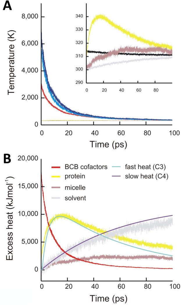
MD simulations of the flow of heat throughout the system. (a) Temperature of the cofactors (dark and light blue, bacteriochlorophylls; red, special pair), protein (yellow), detergent micelle (purple), surrounding solvent (grey) and total system (black) determined from MD trajectories. (b) Heat content of the cofactors (red), protein (yellow), detergent micelle (purple) and solvent (grey). The experimental amplitudes of the non-equilibrated heating (C3, light blue) and equilibrated heating (C4, dark blue) basis spectra are scaled to aid comparison with the heat content of the protein and solvent extracted from MD simulations.

Structural analysis of the protein conformational changes. (a) Theoretical WAXS changes (red) predicted from χ2 fitting to the ultrafast basis spectrum (C1, black) where only movements of the RCvir cofactors were considered. (b) Theoretical WAXS changes (red) predicted from χ2 fitting to the protein basis spectrum (C2, black) where RCvir protein and cofactor atoms were allowed to move. (c) Stereo representation of light induced movements in RCvir. Large spheres represent Cα atoms that display recurrent movements within an ensemble of structural changes selected by χ2 fitting to C2 (Supplementary Fig. 6). Orange spheres represent Cα atoms with recurring movements away from other Cα atoms. No recurrent movements of Cα atoms towards other Cα atoms were identified. Cofactors are shown in grey.
References
-
- Vos MH, Rappaport F, Lambry J-C, Breton J, Martin J-L. Visualization of coherent nuclear motion in a membrane protein by femtosecond spectroscopy. Nature. 1993;363:320–325.
-
- Wang H, et al. Protein dynamics control the kinetics of initial electron transfer in photosynthesis. Science. 2007;316:747–750. - PubMed
Publication types
MeSH terms
Substances
Grants and funding
LinkOut - more resources
Full Text Sources
Other Literature Sources
