. 2014 Sep:62:42-50.
doi: 10.1016/j.mcn.2014.08.003. Epub 2014 Aug 7.

N-myristoylation regulates the axonal distribution of the Fragile X-related protein FXR2P

Affiliations

N-myristoylation regulates the axonal distribution of the Fragile X-related protein FXR2P

Emily E Stackpole et al. Mol Cell Neurosci. 2014 Sep.

Abstract

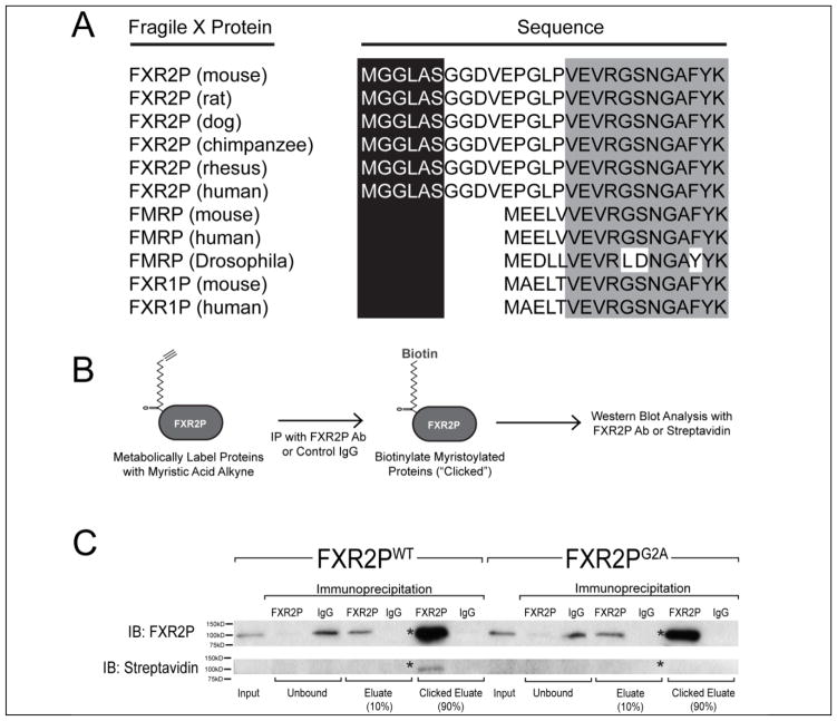
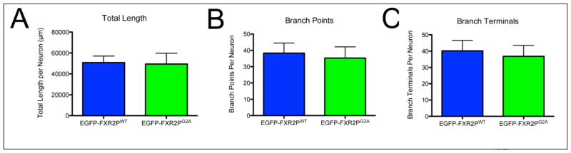
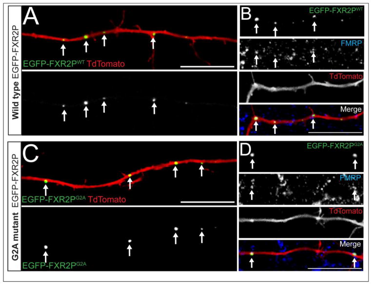
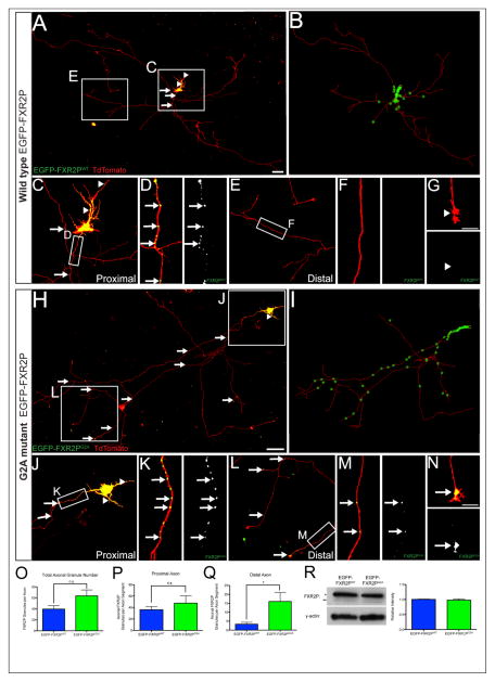
Fragile X syndrome, the leading cause of inherited intellectual disability and autism, is caused by loss of function of Fragile X mental retardation protein (FMRP). FMRP is an RNA binding protein that regulates local protein synthesis in the somatodendritic compartment. However, emerging evidence also indicates important roles for FMRP in axonal and presynaptic functions. In particular, FMRP and its homologue FXR2P localize axonally and presynaptically to discrete endogenous structures in the brain termed Fragile X granules (FXGs). FXR2P is a component of all FXGs and is necessary for the axonal and presynaptic localization of FMRP to these structures. We therefore sought to identify and characterize structural features of FXR2P that regulate its axonal localization. Sequence analysis reveals that FXR2P harbors a consensus N-terminal myristoylation sequence (MGXXXS) that is absent in FMRP. Using click chemistry with wild type and an unmyristoylatable G2A mutant we demonstrate that FXR2P is N-myristoylated on glycine 2, establishing it as a lipid-modified RNA binding protein. To investigate the role of FXR2P N-myristoylation in neurons we generated fluorescently tagged wild type and unmyristoylatable FXR2P (WT and G2A, respectively) and expressed them in primary cortical cultures. Both FXR2P(WT) and FXR2P(G2A) are expressed at equivalent overall levels and are capable of forming FMRP-containing axonal granules. However, FXR2P(WT) granules are largely restricted to proximal axonal segments while granules formed with unmyristoylatable FXR2P(G2A) are localized throughout the axonal arbor, including in growth cones. These studies indicate that N-terminal myristoylation of the RNA binding protein FXR2P regulates its localization within the axonal arbor. Moreover, since FMRP localization within axonal domains requires its association with FXR2P, these findings suggest that FXR2P lipid modification is a control point for the axonal and presynaptic distribution of FMRP.

Keywords: FXR2P; Fragile X syndrome; Myristoylation; RNA binding proteins.


Figures

Figure 1
Figure 1. FXR2P is N-terminally myristoylated
(A) The N-terminus of FXR2P from the six mammalian species analyzed contains a conserved N-terminal myristoylation consensus sequence (MGXXXS). This consensus sequence is not present in either FMRP or FXR1P in any species examined (for clarity, only mouse, human and Drosophila are shown). (B) Schematic of click chemistry-based approach used to detect FXR2P myristoylation (see Methods). (C) Western blot demonstrating N-terminal myristoylation of FXR2PWT but not FXR2PG2A. COS-7 cells transfected with either FXR2PWT or FXR2PG2A were incubated with a biotinylatable analog of myristic acid. Lysates were collected (‘input’) and then immunoprecipitated with either FXR2P antibody or a control IgG. Ninety percent of immunoprecipitates were subjected to the click-iT reaction to biotinylate proteins that had incorporated the myristic analog. Analysis of the western blots with an FXR2P antibody demonstrated the comparable immunoprecipitation of FXR2PWT and FXR2PG2A (asterisk; ~100kD), while neither was detected when a control IgG was used for the immunoprecipitation. Anti-biotin western blotting detected protein that incorporated the myristic analog. FXR2PWT is biotinylated when the click reaction is performed after immunoprecipitation with an FXR2P antibody (asterisk marks ~100kD band corresponding to clicked FXR2P). FXR2PG2A is not biotinylated after immunoprecipitation with an FXR2P antibody and click reaction. Equivalent results were observed in four independent experiments.
Figure 2
Figure 2. Comparable axonal arborization in neurons expressing EGFP-FXR2PWT or EGFP-FXR2PG2A.
TdTomato-filled axonal arbors were reconstructed from neurons co-transfected with either EGFP-FXR2PWT or EGFP-FXR2PG2A. The average total axon length (A), the number of branch points (B) and number of branch terminals (C) in neurons expressing EGFP-FXR2PWT or EGFP-FXR2PG2A was similar. n = 6 reconstructed neurons at DIV6 for each condition.
Figure 3
Figure 3. FXR2P forms granules and associates with FMRP within axons independently of N-myristoylation
DIV3 cortical neuron cultures were co-transfected with TdTomato (red) along with either wild type (A, B) or unmyristoylatable (C, D) EGFP-FXR2P (WT or G2A, respectively; green). Note that both wild type and myristoylation-deficient FXR2P are localized to discrete granules (arrows) in the proximal regions of the axon. Axonal granules containing either EGFP-FXR2PWT (B) or EGFP-FXR2PG2A (D) co-localized with FMRP (blue). DIV6 neurons. Scale bar = 20μm.
Figure 4
Figure 4. Differential axonal distribution of wild type and N-myristoylation-deficient FXR2P
(A) The entire axonal arbor of a cortical neuron expressing wild type FXR2P (EGFP-FXR2PWT; green) and TdTomato (red). EGFP-FXR2PWT localizes to granules in proximal axon segments (arrows). Granules are also observed in dendrites (arrowheads). (B) Neurolucida reconstruction of EGFP-FXR2PWT expressing neuron in A. Green stars mark the location of EGFP-FXR2PWT granules within the axonal arbor (red). (C, D) Progressively higher magnifications showing the abundance of EGFP-FXR2PWT granules (arrows) in proximal axon segments. (EG) In contrast EGFP-FXR2PWT granules were rare in distal axon segments or growth cones. (H) The entire TdTomato filled axonal arbor of a cortical neuron expressing unmyristoylatable FXR2P (EGFP-FXR2PG2A; green). (I) Neurolucida reconstruction of EGFP-FXR2PG2A expressing neuron in G. (JK) Progressively higher magnifications of EGFP-FXR2PG2A localization to numerous granules in proximal axonal segments. (L–M) Progressively higher magnifications showing that EGFP-FXR2PG2A granules are abundant in distal axonal segments. (N) EGFP-FXR2PG2A also localized to growth cones. (O–Q) The distribution of FXR2P granules in axons was analyzed using a Sholl analysis with concentric radii of 20 μm extending from the center of the soma and the number of EGFP-FXR2P puncta within each segment was identified and counted. There was no detectable difference in the total number of granules per axon between EGFP-FXR2PWT and EGFP-FXR2PG2A expressing neurons. At proximal axon segments (<2000 μm from cell soma), there was no difference in the total number of axonal granules between EGFP-FXR2PWT and EGFP-FXR2PG2A expressing neurons. The number of axonal FXR2P granules was significantly increased in distal axonal arbors of neurons expressing EGFP-FXR2PG2A compared to EGFP-FXR2PWT (* = p < 0.05; n = 6 neurons per condition). (R) Western blot analysis using total protein extracts from neuronal cultures expressing either EGFP-FXR2PWT or EGFP-FXR2PG2A indicates that both were expressed at equivalent levels (FXR2P antibody; n = 3 cultures per condition). Upper band in blot is EGFP-FXR2P (*) while lower band is endogenous FXR2P (**). Actin served as a loading control. DIV6 neurons. Scale bar = 100 μm in A, H; 10 μm in G, N.

References

    1. Aderem AA, Albert KA, Keum MM, Wang JK, Greengard P, Cohn ZA. Stimulus- dependent myristoylation of a major substrate for protein kinase C. Nature. 1988;332:362–364. - PubMed
    1. Akins MR, Berk-Rauch HE, Fallon JR. Presynaptic translation: stepping out of the postsynaptic structure. Front Neural Circuits. 2009;3:17. - PMC - PubMed
    1. Akins MR, Leblanc HF, Stackpole EE, Chyung E, Fallon JR. Systematic mapping of fragile X granules in the mouse brain reveals a potential role for presynaptic FMRP in sensorimotor functions. J Comp Neurol. 2012;520:3687–3706. - PMC - PubMed
    1. Antar LN, Li C, Zhang H, Carroll RC, Bassell GJ. Local functions for FMRP in axon growth cone motility and activity-dependent regulation of filopodia and spine synapses. Mol Cell Neurosci. 2006;32:37–48. - PubMed
    1. Bagni C, Greenough WT. From mRNP trafficking to spine dysmorphogenesis: the roots of fragile X syndrome. Nat Rev Neurosci. 2005;6:376–387. - PubMed

Publication types

Substances