. 2015 Jun 4;34(23):2991-9.
doi: 10.1038/onc.2014.243. Epub 2014 Aug 11.

Functional RNAi screen targeting cytokine and growth factor receptors reveals oncorequisite role for interleukin-2 gamma receptor in JAK3-mutation-positive leukemia

Affiliations

Functional RNAi screen targeting cytokine and growth factor receptors reveals oncorequisite role for interleukin-2 gamma receptor in JAK3-mutation-positive leukemia

A Agarwal et al. Oncogene. .

Abstract

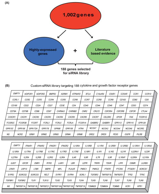
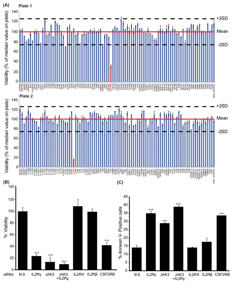
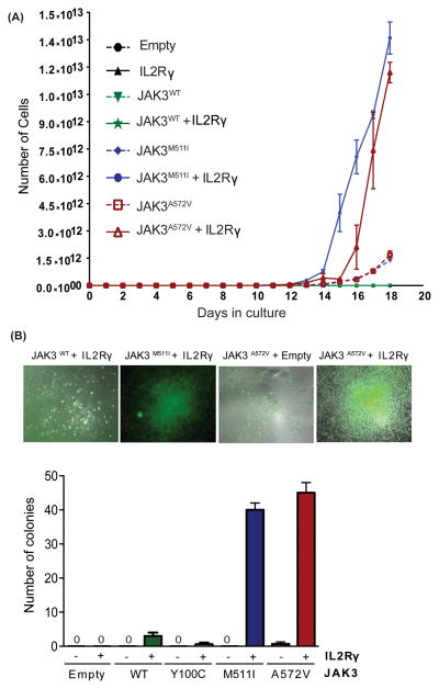
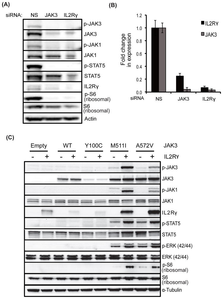
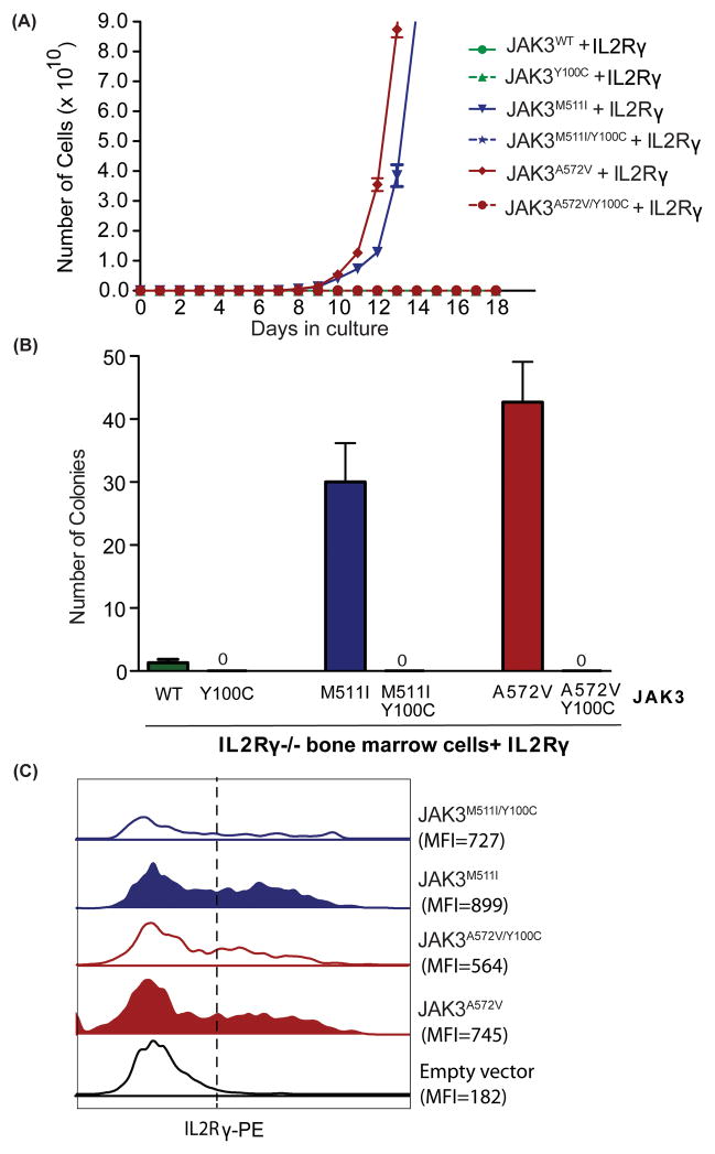
To understand the role of cytokine and growth factor receptor-mediated signaling in leukemia pathogenesis, we designed a functional RNA interference (RNAi) screen targeting 188 cytokine and growth factor receptors that we found highly expressed in primary leukemia specimens. Using this screen, we identified interleukin-2 gamma receptor (IL2Rγ) as a critical growth determinant for a JAK3(A572V) mutation-positive acute myeloid leukemia cell line. We observed that knockdown of IL2Rγ abrogates phosphorylation of JAK3 and downstream signaling molecules, JAK1, STAT5, MAPK and pS6 ribosomal protein. Overexpression of IL2Rγ in murine cells increased the transforming potential of activating JAK3 mutations, whereas absence of IL2Rγ completely abrogated the clonogenic potential of JAK3(A572V), as well as the transforming potential of additional JAK3-activating mutations such as JAK3(M511I). In addition, mutation at the IL2Rγ interaction site in the FERM domain of JAK3 (Y100C) completely abrogated JAK3-mediated leukemic transformation. Mechanistically, we found IL2Rγ contributes to constitutive JAK3 mutant signaling by increasing JAK3 expression and phosphorylation. Conversely, we found that mutant, but not wild-type JAK3, increased the expression of IL2Rγ, indicating IL2Rγ and JAK3 contribute to constitutive JAK/STAT signaling through their reciprocal regulation. Overall, we demonstrate a novel role for IL2Rγ in potentiating oncogenesis in the setting of JAK3-mutation-positive leukemia. In addition, our study highlights an RNAi-based functional assay that can be used to facilitate the identification of non-kinase cytokine and growth factor receptor targets for inhibiting leukemic cell growth.


Conflict of interest statement

CONFLICT OF INTEREST

OHSU and B.J.D. have a financial interest in Molecular MD (5% equity or less). This potential conflict of interest has been reviewed and managed by the OHSU Conflict of Interest in Research Committee and the Integrity Program Oversight Council. OHSU has clinical trial contracts with Novartis, and Bristol Myers Squibb to pay for patient costs, nurse and data manager salaries, and institutional overhead. B.J.D. does not derive salary, nor do their laboratories receive funds, from these contracts. B.J.D serves as a consultant for Roche and Nodality (Consulting income $10,000 or over).

Figures

Figure 1
Figure 1. Functional RNAi screen panel targeting non-kinase cytokine and growth factor receptors
(A) We performed gene expression profiling using mononuclear cells from bone marrow and peripheral blood of 141 leukemia patient samples. This data set was analyzed for 1,002 genes encoding cytokine and growth factor receptors and associated proteins. A total of 188 cancer related genes were selected for inclusion based on the following filtering criteria. First, genes consistently up-regulated compared to the average gene expression of the 100 normalization control probe sets (provided by Affymetrix) in all subjects within each subtype of disease were selected and ranked by average gene expression value. Top 15% genes from all tested subtype of diseases were selected and pooled for inclusion. These genes were designated as highly-expressed genes. Additionally, known cancer-related growth factor genes, based upon evidence found in the literature, were also included. (B) siRNA library comprising 188 genes includes non-kinase cytokine and growth factor receptor families and associated proteins for interleukins, toll-like receptors, tumor necrosis factors, interferons, G-protein coupled receptors, growth hormone receptors and receptors associated with cell differentiation.
Figure 2
Figure 2. Functional RNAi profiling of cytokine and growth factor receptors in CMK cells
(A) CMK cells were electroporated with individual siRNAs targeting 188 genes from cytokine and growth factor receptor families (each siRNA is a pool of four siRNAs). Cells were re-plated in triplicate into culture media for 96 hours, and cell viability was determined by addition of a tetrazolium salt (MTS assay). All values were adjusted to a blank control within the plate and normalized to the median value for all wells on the plate. Effective hits were considered those that robustly decreased cell viability to levels below two standard deviations of the median value for the plate. Bar graph values represent mean percentage (normalized to the median value for all wells on the plate) ± SEM (n= 3). (B, C) Identified targets were confirmed using JAK3 siRNA as a positive control in CMK cells. Effect of gene knock-down on cell viability (B) and apoptosis (C) was measured by MTS assay and Annexin V+ staining, respectively. Values represent mean percentage ± SEM (n= 3). *** denotes p< 0.001.
Figure 3
Figure 3. IL2Rγ is necessary for transformation of JAK3 mutant-positive cells
(A) Ba/F3 cells were retrovirally transfected with the indicated JAK3 mutants and/or IL2Rγ, and sorted using a flow cytometer. Cells were plated in medium lacking WEHI-conditioned medium (source of IL-3), and total viable cells were counted every other day for 18 days. Results shown here are representative of three independent experiments. (B) IL2Rγ−/− Lineage- mouse bone marrow cells were harvested from B6.129S4-Il2rgtm1Wjl/J mice. These cells were co-transfected with retroviruses expressing JAK3 mutants (GFP+) and IL2Rγ. Doubly positive cells (GFP+IL2Rγ+) were FACS sorted and colony formation assays were performed in cytokine-free Methocult media (M3234). Colonies were scored following 8 days of incubation at 37°C in 5% CO2. Values represent mean colony numbers ± SEM (n= 3). Representative colonies are shown.
Figure 4
Figure 4. IL2Rγ regulates JAK3 kinase signaling and reciprocally regulate each other
(A, B) CMK cells were electroporated with JAK3 and IL2Rγ siRNA and cells were cultured for 48 hours. Whole cell lysates were subjected to immunoblotting with the indicated antibodies and actin was used as a loading control (A). Quantitative PCR analysis was performed using specific primers and GAPDH as a reference control (B). (C) Stable Ba/F3 cells were prepared by co-expressing IL2Rγ and JAK3 mutants. The effect of IL2Rγ overexpression on downstream signaling was analyzed by immunobloting with the indicated antibodies and α-tubulin was used as a loading control.
Figure 5
Figure 5. Direct interaction of IL2Rγ and JAK3 is necessary for the transformation of JAK3 mutant-positive cells
(A) Ba/F3 cells were retrovirally transfected with the indicated JAK3 mutants and IL2Rγ, sorted using a flow cytometer. Cells were plated in medium lacking WEHI-conditioned medium (source of IL-3), and total viable cells were counted every other day for 18 days. Results shown here are representative of three independent experiments. (B) IL2Rγ−/− Lineage- mouse bone marrow cells were co-transfected with retroviruses expressing JAK3 mutants (GFP+) and IL2Rγ. Doubly positive cells (GFP+IL2Rγ+) were FACS sorted and colony formation was performed in cytokine-free Methocult media (M3234). Colonies were scored following 8 days of incubation at 37°C in 5% CO2. Values represent mean colony numbers ± SEM (n= 3). (C) Surface expression of IL2Rγ (anti-PE) was analyzed using FACS in JAK3 mutant positive murine bone marrow cells (GFP+) after 48 hrs of co-transfection with retroviruses. Representative histograms showing mean fluorescent intensity is shown.

References

    1. Estey E, Dohner H. Acute myeloid leukaemia. Lancet. 2006 Nov 25;368(9550):1894–907. eng. - PubMed
    1. Graf M, Hecht K, Reif S, Pelka-Fleischer R, Pfister K, Schmetzer H. Expression and prognostic value of hemopoietic cytokine receptors in acute myeloid leukemia (AML): implications for future therapeutical strategies. Eur J Haematol. 2004 Feb;72(2):89–106. eng. - PubMed
    1. Van Etten RA. Aberrant cytokine signaling in leukemia. Oncogene. 2007 Oct 15;26(47):6738–49. - PMC - PubMed
    1. Vainchenker W, Constantinescu SN. JAK/STAT signaling in hematological malignancies. Oncogene. 2013 May 23;32(21):2601–13. - PubMed
    1. Tefferi A. Novel mutations and their functional and clinical relevance in myeloproliferative neoplasms: JAK2, MPL, TET2, ASXL1, CBL, IDH and IKZF1. Leukemia : official journal of the Leukemia Society of America, Leukemia Research Fund, UK. 2010 Jun;24(6):1128–38. eng. - PMC - PubMed

Publication types

MeSH terms

Associated data