. 2015 Jun 11;34(24):3199-206.
doi: 10.1038/onc.2014.242. Epub 2014 Aug 11.

Oncogenic suppression of apoptosis uncovers a Rac1/JNK proliferation pathway activated by loss of Par3

Affiliations

Oncogenic suppression of apoptosis uncovers a Rac1/JNK proliferation pathway activated by loss of Par3

A Archibald et al. Oncogene. .

Abstract

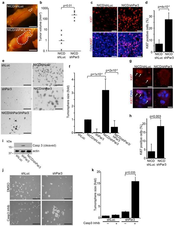
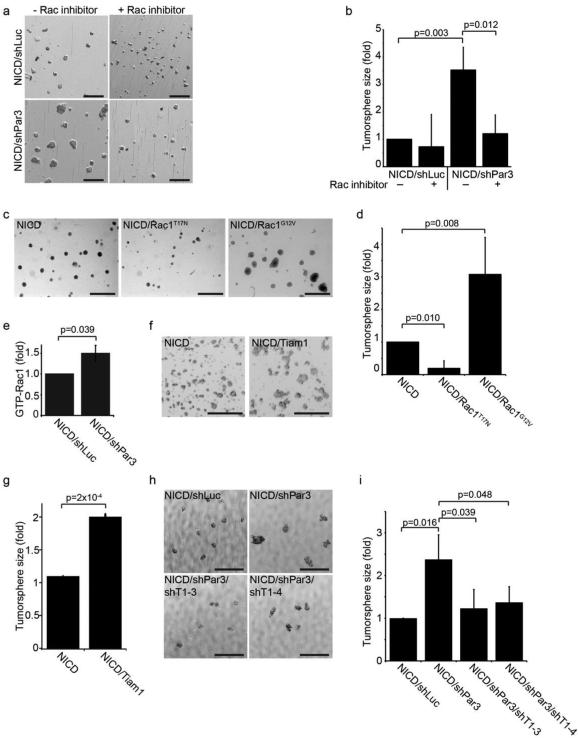
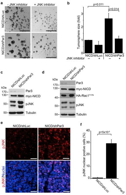
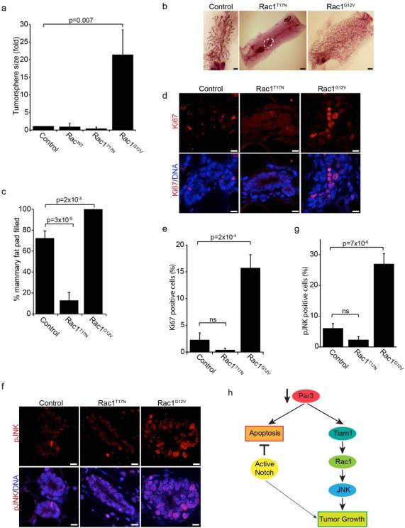
Disruption of epithelial organization and loss of growth control are universal features of carcinomas, yet how these features are linked during cancer progression remains poorly understood. Cell polarity proteins control cellular and tissue organization and are emerging as important mediators of cancer progression. The Par3 polarity protein is a molecular scaffold that functions to recruit and spatially organize signaling factors, and was recently identified as a suppressor of breast cancer invasion and metastasis. Here, we show that loss of Par3 in mammary epithelial cells promotes apoptosis, and that oncogenic Notch overcomes the apoptotic signal to reveal an unexpected pro-proliferative role for loss of Par3 in mammary tumors. In this context, loss of Par3 deregulates Rac1 activity to activate Jun N-terminal Kinase-dependent proliferation and tumor growth. Thus, we demonstrate a mechanism by which loss of Par3 promotes proliferation and tumorigenesis, which supports a tumor-suppressive function for Par3 in the mammary epithelium.


Figures

Figure 1
Figure 1. Loss of Par3 cooperates with NICD to promote mammary tumor growth
(a) Mammary tumors from mammary epithelial cells expressing NICD/shLuc (control) or NICD/shPar3 (shRNA sequence: shPar3 5’-acaagcgtggcatgatcca). Primary mammary epithelial cells (MECs) were isolated from wild-type mice, infected with lentivirus to express NICD and shRNA, then transplanted into the cleared mammary fat pad. (b) Volumes of NICD/shLuc and NICD/shPar3 tumors were calculated using the formula V=□/6×(length×width). (c) Tumor sections were immunostained for Ki67 (1:100, Abcam). (d) Quantification of cells with nuclear Ki67 from (c). Two regions from each of 3 tumors were analyzed for each. (e) MECs were infected with lentivirus to express shLuc, NICD/shLuc, shPar3, NICD/shPar3, or NICD/shPar3/hPar3 and were grown in suspension cultures for 9 days to form tumorspheres. hPar3 is resistant to the mouse-specific Par3 shRNA. (f) Quantification of tumorsphere sizes from (d). The area of each tumorsphere was measured using a custom ImageJ macro. Each independent experiment contained at least 30 tumorspheres. (g) Tumorspheres were grown for 4 days, then immunostained for Ki67. Arrows show Ki67 positive cells. (h) Quantification of cells with nuclear Ki67 from (g); n= 4 independent experiments. (i) Cell lysates were prepared from tumorspheres and immunoblotted for cleaved Caspase-3 (1:1000, Cell Signaling Technology) or actin (1:1000, Sigma) as a loading control. (j) MECs expressing shLuc or shPar3 were treated with DMSO (control) or 10μM Caspase-3 inhibitor (Ac-DEVD-CHO, BD Biosciences), and were grown for 9 days in suspension culture. (k) Quantification of tumorspheres in (j). n=3 independent samples, unless otherwise noted. Error bars=sd. p-values were calculated using the student’s t-test (b, d, h) or the ANOVA with Tukey HSD post-hoc test (f, k) using SPSS software. Scale bars=1 cm (a); 20μm (c); 1 mm (e); 200μm (j).
Figure 2
Figure 2. Loss of Par3 promotes tumorsphere growth through Rac1 activation by Tiam1
(a) Tumorsphere cultures of MECs expressing NICD/shLuc or NICD/shPar3 in the presence or absence of 25 μM Rac1 inhibitor (NSC23766, Tocris). (b) Quantification of tumorspheres sizes from (a). (c) Tumorsphere cultures of MECs expressing NICD, NICD/Rac1T17N (dominant negative), or NICD/Rac1G12V (constitutively active). (d) Quantification of tumorsphere sizes from (c). (e) Levels of total active Rac1-GTP were measured in NICD/shLuc and NICD/shPar3 tumorspheres using a GLISA assay kit (Cat. # BK128, Cytoskeleton Inc.), n=5. (f) Tumorsphere cultures of MECs expressing NICD or NICD/Tiam1. (g) Quantification of tumorsphere sizes from (f). (h) Tumorspheres of MECs expressing NICD/shLuc or NICD/shPar3 and one of two Tiam1 shRNA (shRNA sequences: shTiam1-3 5’-cgatgactttatatttata; shTiam1-4 5’-tttcgtgctatgatgaatc). (i) Quantification of tumorsphere sizes in (h), n=4. n=3 independent samples, unless otherwise noted. p-values were calculated using the ANOVA with Tukey HSD post-hoc test (b, d, i) or the student’s t-test (e, g). Error bars represent sd. Scale bars=1mm.
Figure 3
Figure 3. Par3 regulates tumor growth by activating JNK
(a) Tumorsphere cultures of MECs expressing NICD/shLuc or NICD/shPar3 in the presence or absence of 25μM JNK inhibitor (SP600125, Sigma). (b) Quantification of tumorsphere sizes from (a), n=3. (c and d) Immunoblots of NICD/shLuc and NICD/shPar3 tumorsphere lysates (c) or co-expressing dominant negative HA-Rac1T17N (d). Antibodies used were rabbit anti-Par3 (Macara lab12), mouse anti-Myc (1:1000, McGill hybridoma facility), rabbit anti-HA (1:1000, McGill hybridoma facility), rabbit anti-pJNK (pThr183/Tyr185, 1:1000, Cell Signaling Technology), mouse anti-a-tubulin as a loading control (1:5000, Sigma). (e) NICD/shLuc and NICD/shPar3 mammary tumor sections were stained for phospho-JNK (1:100). (f) Quantification of cells with nuclear phospho-JNK from (e). Two regions from each of 3 tumors were analyzed for each. p-values were calculated using the ANOVA with Tukey HSD post-hoc test (b) or student’s t-test (f). Error bars represent sd (e) or sem (f). Scale bars = 1mm (a) and 20μm (e).
Figure 4
Figure 4. Rac1/JNK signaling regulates mammary epithelial proliferation
(a) Quantification of tumorsphere sizes of MECs expressing GFP (control), wild-type (Rac1WT), dominant negative (Rac1T17N) or constitutively active (Rac1G12V) Rac1. (b) Mammary epithelial cells were transduced with control (GFP alone), Rac1T17N, or Rac1G12V lentivirus and then transplanted into a cleared mammary fat pad and allowed to regrow for 8-weeks. (c) Quantification of the percentage of the fat pad filled from (b), n=4. (d) Tissue sections of control (GFP), Rac1T17N, and Rac1G12V mammary glands were immunostained for Ki67. (e) Quantification of cells with nuclear Ki67 from (d). All ducts from 2 tissue sections for each of 3 glands were measured. (f) Tissue sections of control (GFP), Rac1T17N, and Rac1G12V mammary glands were stained for phospho-JNK. (g) Quantification of cells with nuclear phospho-JNK from (f). (h) Model showing that loss of Par3 in the normal mammary epithelium activates both apoptotic and proliferative responses. In the presence of an anti-apoptotic factor (i.e. Notch oncogene), the apoptotic response caused by a loss of Par3 is suppressed and a Tiam1/Rac1/JNK mediated proliferative response is revealed. p-values were calculated using the ANOVA with Tukey HSD post-hoc test. Error bars represent sd (a) or sem (c, e, and (g). Scale bars = 2mm (b) or 10 μm (d and f).

References

    1. McCaffrey LM, Macara IG. Epithelial organization, cell polarity and tumorigenesis. Trends Cell Biol. 2011 Dec;21(12):727–35. - PubMed
    1. Tepass U. The apical polarity protein network in Drosophila epithelial cells: regulation of polarity, junctions, morphogenesis, cell growth, and survival. Annu Rev Cell Dev Biol. 2012;28:655–85. - PubMed
    1. Zhao B, Tumaneng K, Guan KL. The Hippo pathway in organ size control, tissue regeneration and stem cell self-renewal. Nat Cell Biol. 2011 Aug;13(8):877–83. - PMC - PubMed
    1. Huang L, Muthuswamy SK. Polarity protein alterations in carcinoma: a focus on emerging roles for polarity regulators. Curr Opin Genet Dev. 2010 Feb;20(1):41–50. PubMed PMID: 20093003. - PMC - PubMed
    1. Hayase J, Kamakura S, Iwakiri Y, Yamaguchi Y, Izaki T, Ito T, et al. The WD40 protein Morg1 facilitates Par6-aPKC binding to Crb3 for apical identity in epithelial cells. J Cell Biol. 2013 Mar 4;200(5):635–50. - PMC - PubMed

Publication types

MeSH terms