Roxithromycin treatment inhibits TGF-β1-induced activation of ERK and AKT and down-regulation of caveolin-1 in rat airway smooth muscle cells
- PMID: 25109503
- PMCID: PMC4256937
- DOI: 10.1186/s12931-014-0096-z
Roxithromycin treatment inhibits TGF-β1-induced activation of ERK and AKT and down-regulation of caveolin-1 in rat airway smooth muscle cells
Abstract
Background: Roxithromycin (RXM) has been widely used in asthma treatment; however, the mechanism has not been fully understood. The aim of our study was to investigate the underlying mechanism of RXM treatment in mediating the effect of transforming growth factor (TGF)-β1 on airway smooth muscle cells (ASMCs) proliferation and caveolinn-1 expression.
Methods: Firstly, the rat ovalbumin (OVA) model was built according to the previous papers. Rat ASMCs were prepared and cultured, and then TGF-β1 production in ASMCs was measured by enzyme-linked immunosorbent assay (ELISA). Moreover, the proliferation of ASMCs was determined using cell counting kit (CCK-8) assay. Additionally, the expressions of caveolin-1, phosphorylated-ERK1/2 (p-ERK1/2) and phosphorylated-AKT (p-AKT) in ASMCs treated with or without PD98059 (an ERK1/2 inhibitor), wortannin (a PI3K inhibitor), β-cyclodextrin (β-CD) and RXM were measured by Western blot. Finally, data were evaluated using t-test or one-way ANOVA, and then a P value < 0.05 was set as a threshold.
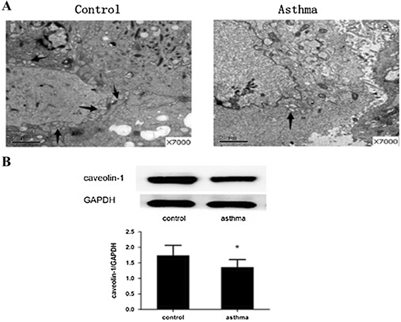
Results: Compared with normal control, TGF-β1 secretion was significantly increased in asthmatic ASMCs; meanwhile, TGF-β1 promoted ASMCs proliferation (P < 0.05). However, ASMCs proliferation was remarkably inhibited by RXM, β-CD, PD98059 and wortmannin (P < 0.05). Moreover, the expressions of p-ERK1/2 and p-AKT were increased and peaked at 20 min after TGF-β1 stimulation, and then suppressed by RXM. Further, caveolin-1 level was down-regulated by TGF-β1 and up-regulated by inhibitors and RXM.
Conclusion: Our findings demonstrate that RXM treatment inhibits TGF-β1-induced activation of ERK and AKT and down-regulation of caveolin-1, which may be the potential mechanism of RXM protection from chronic inflammatory diseases, including bronchial asthma.
Figures


Similar articles
-
Roxithromycin inhibits VEGF-induced human airway smooth muscle cell proliferation: Opportunities for the treatment of asthma.Exp Cell Res. 2016 Oct 1;347(2):378-84. doi: 10.1016/j.yexcr.2016.08.024. Epub 2016 Aug 29. Exp Cell Res. 2016. PMID: 27587274
-
[Functional role of caveolin-1 in airway smooth muscle cells proliferation and regulatory effect of roxithromycin].Zhonghua Yi Xue Za Zhi. 2013 Sep 10;93(34):2750-4. Zhonghua Yi Xue Za Zhi. 2013. PMID: 24360114 Chinese.
-
MiRNA-620 promotes TGF-β1-induced proliferation of airway smooth muscle cell through controlling PTEN/AKT signaling pathway.Kaohsiung J Med Sci. 2020 Nov;36(11):869-877. doi: 10.1002/kjm2.12260. Epub 2020 Jun 24. Kaohsiung J Med Sci. 2020. PMID: 32583575 Free PMC article.
-
Roxithromycin suppresses airway remodeling and modulates the expression of caveolin-1 and phospho-p42/p44MAPK in asthmatic rats.Int Immunopharmacol. 2015 Feb;24(2):247-255. doi: 10.1016/j.intimp.2014.11.015. Epub 2014 Nov 25. Int Immunopharmacol. 2015. PMID: 25479721
-
MiR-328-3p promotes TGF-β1-induced proliferation, migration, and inflammation of airway smooth muscle cells by regulating the PTEN/Akt pathway.Allergol Immunopathol (Madr). 2023 Mar 1;51(2):151-159. doi: 10.15586/aei.v51i2.767. eCollection 2023. Allergol Immunopathol (Madr). 2023. PMID: 36916101
Cited by
-
Azithromycin induces apoptosis in airway smooth muscle cells through mitochondrial pathway in a rat asthma model.Ann Transl Med. 2021 Jul;9(14):1181. doi: 10.21037/atm-21-3478. Ann Transl Med. 2021. PMID: 34430622 Free PMC article.
-
Mesenchymal stem cells suppress lung inflammation and airway remodeling in chronic asthma rat model via PI3K/Akt signaling pathway.Int J Clin Exp Pathol. 2015 Aug 1;8(8):8958-67. eCollection 2015. Int J Clin Exp Pathol. 2015. PMID: 26464637 Free PMC article.
-
The critical roles of caveolin-1 in lung diseases.Front Pharmacol. 2024 Sep 24;15:1417834. doi: 10.3389/fphar.2024.1417834. eCollection 2024. Front Pharmacol. 2024. PMID: 39380904 Free PMC article. Review.
-
IL-38 alleviates airway remodeling in chronic asthma via blocking the profibrotic effect of IL-36γ.Clin Exp Immunol. 2023 Dec 13;214(3):260-274. doi: 10.1093/cei/uxad099. Clin Exp Immunol. 2023. PMID: 37586814 Free PMC article.
-
Mitochondrial ATP-Sensitive K+ Channel Opening Increased the Airway Smooth Muscle Cell Proliferation by Activating the PI3K/AKT Signaling Pathway in a Rat Model of Asthma.Can Respir J. 2021 Jul 12;2021:8899878. doi: 10.1155/2021/8899878. eCollection 2021. Can Respir J. 2021. PMID: 34336047 Free PMC article.
References
-
- Ito I, Fixman ED, Asai K, Yoshida M, Gounni AS, Martin JG, Hamid Q. Platelet-derived growth factor and transforming growth factor-beta modulate the expression of matrix metalloproteinases and migratory function of human airway smooth muscle cells. Clin Exp Allergy. 2009;39(9):1370–1380. doi: 10.1111/j.1365-2222.2009.03293.x. - DOI - PubMed
Publication types
MeSH terms
Substances
LinkOut - more resources
Full Text Sources
Other Literature Sources
Miscellaneous

