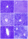
Cytoprotective effects of amifostine, ascorbic acid and N-acetylcysteine against methotrexate-induced hepatotoxicity in rats
- PMID: 25110444
- PMCID: PMC4123346
- DOI: 10.3748/wjg.v20.i29.10158
Cytoprotective effects of amifostine, ascorbic acid and N-acetylcysteine against methotrexate-induced hepatotoxicity in rats
Abstract
Aim: To investigate the potential role of oxidative stress and the possible therapeutic effects of N-acetyl cysteine (NAC), amifostine (AMF) and ascorbic acid (ASC) in methotrexate (MTX)-induced hepatotoxicity.
Methods: An MTX-induced hepatotoxicity model was established in 44 male Sprague Dawley rats by administration of a single intraperitoneal injection of 20 mg/kg MTX. Eleven of the rats were left untreated (Model group; n = 11), and the remaining rats were treated with a 7-d course of 50 mg/kg per day NAC (MTX + NAC group; n = 11), 50 mg/kg per single dose AMF (MTX + AMF group; n = 11), or 10 mg/kg per day ASC (MTX + ASC group; n = 11). Eleven rats that received no MTX and no treatments served as the negative control group. Structural and functional changes related to MTX- and the various treatments were assessed by histopathological analysis of liver tissues and biochemical assays of malondialdehyde (MDA), superoxide dismutase (SOD), catalase, glutathione (GSH) and xanthine oxidase activities and of serum levels of aspartate aminotransferase, alanine aminotransferase, alkaline phosphatase and total bilirubin.
Results: Exposure to MTX caused structural and functional hepatotoxicity, as evidenced by significantly worse histopathological scores [median (range) injury score: control group: 1 (0-3) vs 7 (6-9), P = 0.001] and significantly higher MDA activity [409 (352-466) nmol/g vs 455.5 (419-516) nmol/g, P < 0.05]. The extent of MTX-induced perturbation of both parameters was reduced by all three cytoprotective agents, but only the reduction in hepatotoxicity scores reached statistical significance [4 (3-6) for NAC, 4.5 (3-5) for AMF and 6 (5-6) for ASC; P = 0.001, P = 0.001 and P < 0.005 vs model group respectively]. Exposure to MTX also caused a significant reduction in the activities of GSH and SOD antioxidants in liver tissues [control group: 3.02 (2.85-3.43) μmol/g and 71.78 (61.88-97.81) U/g vs model group: 2.52 (2.07-3.34) μmol/g and 61.46 (58.27-67.75) U/g, P < 0.05]; however, only the NAC treatment provided significant increases in these antioxidant enzyme activities [3.22 (2.54-3.62) μmol/g and 69.22 (61.13-100.88) U/g, P < 0.05 and P < 0.01 vs model group respectively].
Conclusion: MTX-induced structural and functional damage to hepatic tissues in rats may involve oxidative stress, and cytoprotective agents (NAC > AMF > ASC) may alleviate MTX hepatotoxicity.
Keywords: Amifostine; Ascorbic acid; Hepatotoxicity; Methotrexate; N-acetyl cysteine; Oxidative stress.
Figures


References
-
- Soliman ME. Evaluation of the possible protective role of folic acid on the liver toxicity ınduced experimentally by methotrexate in adult male albino rats. Egypt J Histol. 2009;32:118–128.
-
- Dalaklioglu S, Genc GE, Aksoy NH, Akcit F, Gumuslu S. Resveratrol ameliorates methotrexate-induced hepatotoxicity in rats via inhibition of lipid peroxidation. Hum Exp Toxicol. 2013;32:662–671. - PubMed
-
- Cetin A, Kaynar L, Eser B, Karada C, Saraymen B, Öztürk A. Beneficial effects of propolis on methotrexate-induced liver ınjury in rats. Acta Oncologica Turcica. 2011;44:18–23.
-
- Vardi N, Parlakpinar H, Cetin A, Erdogan A, Cetin Ozturk I. Protective effect of beta-carotene on methotrexate-induced oxidative liver damage. Toxicol Pathol. 2010;38:592–597. - PubMed
Publication types
MeSH terms
Substances
LinkOut - more resources
Full Text Sources
Other Literature Sources
Medical
Miscellaneous

