Review
. 2014:2014:783289.
doi: 10.1155/2014/783289. Epub 2014 Jul 3.

The extracellular matrix contributes to mechanotransduction in uterine fibroids

Affiliations
Review

The extracellular matrix contributes to mechanotransduction in uterine fibroids

Phyllis C Leppert et al. Obstet Gynecol Int. 2014.

Abstract

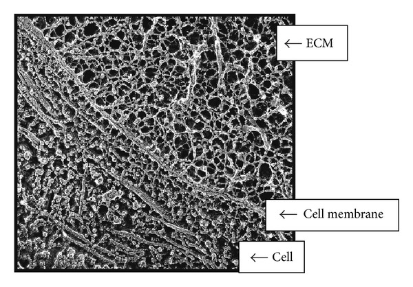
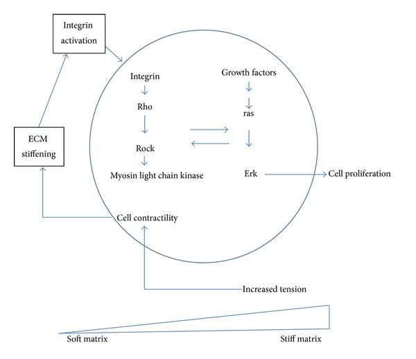
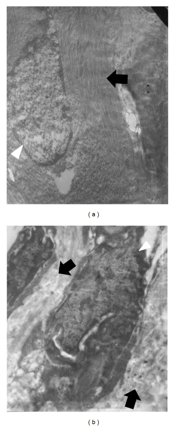
The role of the extracellular matrix (ECM) and mechanotransduction as an important signaling factor in the human uterus is just beginning to be appreciated. The ECM is not only the substance that surrounds cells, but ECM stiffness will either compress cells or stretch them resulting in signals converted into chemical changes within the cell, depending on the amount of collagen, cross-linking, and hydration, as well as other ECM components. In this review we present evidence that the stiffness of fibroid tissue has a direct effect on the growth of the tumor through the induction of fibrosis. Fibrosis has two characteristics: (1) resistance to apoptosis leading to the persistence of cells and (2) secretion of collagen and other components of the ECM such a proteoglycans by those cells leading to abundant disposition of highly cross-linked, disoriented, and often widely dispersed collagen fibrils. Fibrosis affects cell growth by mechanotransduction, the dynamic signaling system whereby mechanical forces initiate chemical signaling in cells. Data indicate that the structurally disordered and abnormally formed ECM of uterine fibroids contributes to fibroid formation and growth. An appreciation of the critical role of ECM stiffness to fibroid growth may lead to new strategies for treatment of this common disease.


Figures

Figure 1
Figure 1
Interconnected structural components of cells and ECM. Quick freeze, deep etch electron micrograph of a fetal ear cartilage chondrocyte cell to illustrate the integration of structures inside and outside the cell. The ECM (containing a meshwork of proteoglycans, collagen, fibronectin, and laminin), the cell membrane (integrins receptors), and the cell (containing microtubules) are visualized. In the ECM the thinnest fibrils in the meshwork are 4 ± I nm and are presumed to be proteoglycans. The larger thicker fibrils are collagen. Micrograph courtesy of Robert Mecham and John Heuser, Washington University, St. Louis, MO, USA. This figure illustrates that mechanical forces can be transmitted across the cell surface and into the cell by means of interconnected structural components (as discussed in [4]).
Figure 2
Figure 2
Elements of mechanical signaling. A simplified pathway of mechanical signaling in cells is depicted. As the cell cytoskeleton contracts (a process called cell contractility) and ECM accumulates in the cell microenvironment, integrin activation occurs leading to activation of Rho (as discussed in [1]). Rho in turn activates ROCK leading to activation of ras. In fibroid cells obtained either at the time of hysterectomy or myomectomy, RhoA activity is attenuated. This adaptation of the fibroid cell is not ROCK dependent (as discussed in [2]). These findings suggest that fibroid cells in symptomatic tumors where treatment was needed are fundamentally adapted to their stiff microenvironment and thus become insensitive to moderate mechanical cues. It is not certain when in the natural history of fibroid development that this adaptive response to mechanotransduction occurs. Nevertheless, mechanical sensing does occur in fibroid cells.
Figure 3
Figure 3
Collagen fibrils in myometrium and fibroids. Comparison of collagen fibril organization in the extracellular matrix of myometrium or uterine fibroid using electron microscopy. (a) Myometrium. Collagen fibrils are tightly packed and well-aligned, as shown by the black arrow. The nucleus is denoted by the white arrowhead. Magnification = 11,500x. (b) Fibroid. The collagen fibrils are randomly aligned and widely spaced, as shown by black arrows. The nucleus is notched and denoted by the white arrowhead. Magnification = 15,500x. Representative sections on samples harvested from a single uterus.

References

    1. Rogers R, Norian J, Malik M, et al. Mechanical homeostasis is altered in uterine leiomyoma. The American Journal of Obstetrics and Gynecology. 2008;198(4):474.e1–474.e11. - PMC - PubMed
    1. Norian JM, Owen CM, Taboas J, et al. Characterization of tissue biomechanics and mechanical signaling in uterine leiomyoma. Matrix Biology. 2012;31(1):57–65. - PMC - PubMed
    1. Jayes FL, Ma X, Flannery EM, Moutos FT, Guilak F, Leppert PC. Biomechanical Evaluation of Human Uterine Fibroids after Exposure to Purified Clostridial Collagenase. Montreal, Canada: Society for the Study of Reproduction; 2013.
    1. Wang N, Butler JP, Ingber DE. Mechanotransduction across the cell surface and through the cytoskeleton. Science. 1993;260(5111):1124–1127. - PubMed
    1. Jaalouk DE, Lammerding J. Mechanotransduction gone awry. Nature Reviews Molecular Cell Biology. 2009;10(1):63–73. - PMC - PubMed