. 2014 Oct;87(1042):20140311.
doi: 10.1259/bjr.20140311. Epub 2014 Aug 11.

Development of an applicator for eye lens dosimetry during radiotherapy

Affiliations

Development of an applicator for eye lens dosimetry during radiotherapy

J M Park et al. Br J Radiol. 2014 Oct.

Abstract

Objective: To develop an applicator for in vivo measurements of lens dose during radiotherapy.

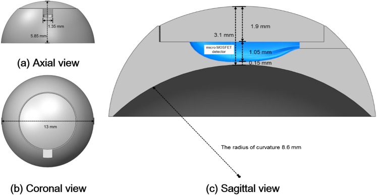
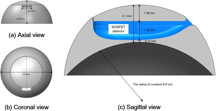
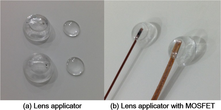
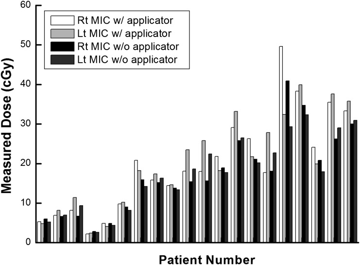
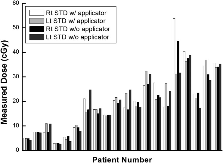
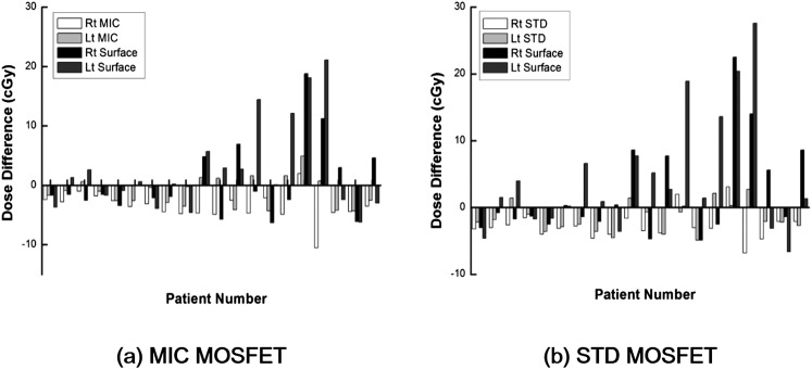
Methods: A contact lens-shaped applicator made of acrylic was developed for in vivo measurements of lens dose. This lens applicator allows the insertion of commercially available metal oxide semiconductor field effect transistors (MOSFETs) dosemeters. CT images of an anthropomorphic phantom with and without the applicator were acquired. Ten volumetric modulated arc therapy plans each for the brain and the head and neck cancer were generated and delivered to an anthropomorphic phantom. The differences between the measured and the calculated doses at the lens applicator, as well as the differences between the measured and the calculated doses at the surface of the eyelid were acquired.

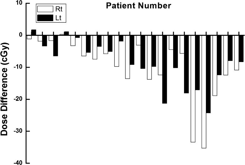
Results: The average difference between the measured and the calculated doses with the applicator was 3.1 ± 1.8 cGy with a micro MOSFET and 2.8 ± 1.3 cGy with a standard MOSFET. The average difference without the lens applicator was 4.8 ± 5.2 cGy with the micro MOSFET and 5.7 ± 6.5 cGy with the standard MOSFET. The maximum difference with the micro MOSFET was 10.5 cGy with the applicator and 21.1 cGy without the applicator. For the standard MOSFET, it was 6.8 cGy with the applicator and 27.6 cGy without the applicator.

Conclusion: The lens applicator allowed reduction of the differences between the calculated and the measured doses during in vivo measurement for the lens compared with in vivo measurement at the surface of the eyelid.

Advances in knowledge: By using an applicator for in vivo dosimetry of the eye lens, it was possible to reduce the measurement uncertainty.


Figures

Figure 1.
Figure 1.
The axial (a), coronal (b) and sagittal (c) views of the lens applicator for in vivo dosimetry of lens dose using micro metal oxide semiconductor field effect transistor (MOSFET) dosemeter are shown. The diameter and the height were 13.00  and 5.85 mm, respectively. The thickness from the dosemeter to the ocular surface was 0.15 mm. The applicator was made of acrylic and consisted two parts that were a cover and a base.
Figure 2.
Figure 2.
The axial (a), coronal (b) and sagittal (c) views of the lens applicator for in vivo dosimetry of lens dose using standard metal oxide semiconductor field effect transistor (MOSFET) dosemeter are shown. The diameter and the height were 13.00  and 5.85 mm, respectively. The thickness from the dosemeter to the ocular surface was 0.15 mm. The applicator was made of acrylic and consisted two parts that were a cover and a base.
Figure 3.
Figure 3.
Two-types of lens applicators for lens in in vivo dosimetry are shown (a). One was for the insertion of micro metal oxide semiconductor field effect transistor (MOSFET) dosemeter (top) and the other was for the insertion of standard MOSFET dosemeter (bottom). Micro and standard MOSFET dosemeter inserted into the lens applicators are shown (b).
Figure 4.
Figure 4.
The dose linearity (a), dose rate dependency (b), depth dose dependency (c) and angular dependency (d) of both micro (MIC) and standard (STD) metal oxide semiconductor field effect transistors (MOSFET) dosemeter are shown. The percent depth doses (PDDs) acquired with MOSFET dosemeter were compared with the PDDs acquired with parallel-plate chamber. The measured values with MOSFET dosemeter at various gantry angles were compared with the measured value at the gantry angle of 0°. IC, ion chamber; MU, monitor unit.
Figure 5.
Figure 5.
The measured values using micro metal oxide semiconductor field effect transistor (MOSFET) dosemeter at the surface of eyelid and at the lens applicator are shown in units of centigray. Lt, left; MIC, micro; Rt, right; w/, with; w/o, without.
Figure 6.
Figure 6.
The measured values using metal oxide semiconductor field effect transistors dosemeter at the surface of eyelid and at the lens applicator are shown in units of centigray. Lt, left; Rt, right; STD, standard; w/, with; w/o, without.
Figure 7.
Figure 7.
The differences between the measured values with standard metal oxide semiconductor field effect transistor dosemeter at the surface of the eyelid and the calculated dose to the lens with treatment planning system for each patient are shown in units of centigray. Lt, left; Rt, right.
Figure 8.
Figure 8.
The differences between the measured dose with micro metal oxide semiconductor field effect transistor (MOSFET) dosemeter [micro (MIC)] (a) inserted into the lens applicator and the calculated dose at the lens applicator with treatment planning system as well as the differences between the measured dose at the surface of eyelid and the calculated dose at the same location are shown in units of centigray for each patient. The same data with standard MOSFET dosemeter are also shown (b). Lt, left; Rt, right; STD, standard.

References

    1. Henk JM, Whitelocke RA, Warrington AP, Bessell EM. Radiation dose to the lens and cataract formation. Int J Radiat Oncol Biol Phys 1993; 25: 815–20. - PubMed
    1. Pawlicki T, Luxton G, Le QT, Findley D, Ma CM. Lens dose in MLC-based IMRT treatments of the head and neck. Int J Radiat Oncol Biol Phys 2004; 59: 293–9. doi: 10.1016/j.ijrobp.2004.01.019 - DOI - PubMed
    1. Woo SY, Donaldson SS, Heck RJ, Nielson KL, Shostak C. Minimizing and measuring lens dose when giving cranial irradiation. Radiother Oncol 1989; 16: 183–8. - PubMed
    1. Ma L, Chin L, Sarfaraz M, Shepard D, Yu C. An investigation of eye lens dose for gamma knife treatments of trigeminal neuralgia. J Appl Clin Med Phys 2000; 1: 116–19. doi: 10.1120/1.1308191 - DOI - PMC - PubMed
    1. Behrens R, Dietze G. Dose conversion coefficients for photon exposure of the human eye lens. Phys Med Biol 2011; 56: 415–37. doi: 10.1088/0031-9155/56/2/009 - DOI - PubMed

Publication types