Review
. 2015 Jan;56(1):1-21.
doi: 10.1002/em.21892. Epub 2014 Aug 11.

Recognition and repair of chemically heterogeneous structures at DNA ends

Affiliations
Review

Recognition and repair of chemically heterogeneous structures at DNA ends

Sara N Andres et al. Environ Mol Mutagen. 2015 Jan.

Abstract

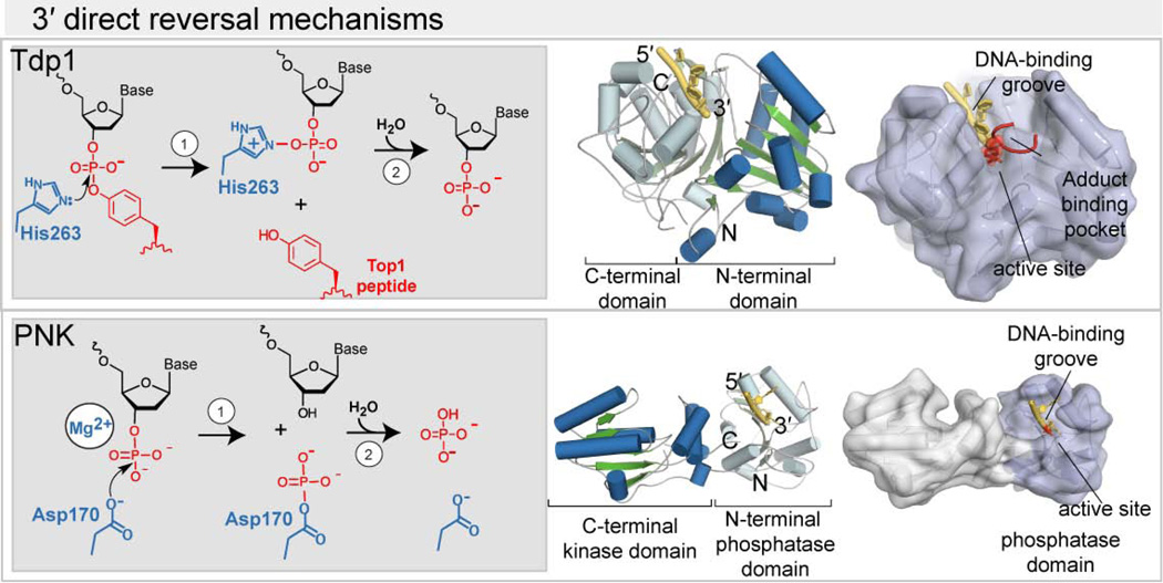
Exposure to environmental toxicants and stressors, radiation, pharmaceutical drugs, inflammation, cellular respiration, and routine DNA metabolism all lead to the production of cytotoxic DNA strand breaks. Akin to splintered wood, DNA breaks are not "clean." Rather, DNA breaks typically lack DNA 5'-phosphate and 3'-hydroxyl moieties required for DNA synthesis and DNA ligation. Failure to resolve damage at DNA ends can lead to abnormal DNA replication and repair, and is associated with genomic instability, mutagenesis, neurological disease, ageing and carcinogenesis. An array of chemically heterogeneous DNA termini arises from spontaneously generated DNA single-strand and double-strand breaks (SSBs and DSBs), and also from normal and/or inappropriate DNA metabolism by DNA polymerases, DNA ligases and topoisomerases. As a front line of defense to these genotoxic insults, eukaryotic cells have accrued an arsenal of enzymatic first responders that bind and protect damaged DNA termini, and enzymatically tailor DNA ends for DNA repair synthesis and ligation. These nucleic acid transactions employ direct damage reversal enzymes including Aprataxin (APTX), Polynucleotide kinase phosphatase (PNK), the tyrosyl DNA phosphodiesterases (TDP1 and TDP2), the Ku70/80 complex and DNA polymerase β (POLβ). Nucleolytic processing enzymes such as the MRE11/RAD50/NBS1/CtIP complex, Flap endonuclease (FEN1) and the apurinic endonucleases (APE1 and APE2) also act in the chemical "cleansing" of DNA breaks to prevent genomic instability and disease, and promote progression of DNA- and RNA-DNA damage response (DDR and RDDR) pathways. Here, we provide an overview of cellular first responders dedicated to the detection and repair of abnormal DNA termini.

Keywords: DNA damage response; X-ray crystallography; cancer; neurodegeneration.


Figures

Figure 1
Figure 1. Chemically heterogeneous, “dirty”, DNA end structures
Repair of strand breaks by DNA ligases requires 5′–phosphate and 3′–hydroxyl groups. Dirty ends have chemical modifications (red) at either the 5′– or 3′–ends that will interfere with ligation. Sources of specific types of damage and enzymes that can repair them are indicated.
Figure 2
Figure 2
Map of DNA end cleansing pathways. Networks of end-cleansing proteins repair specific types of end damage before DNA ligase can complete the final break–sealing reaction. Unrepaired DNA damage can interfere with the function of enzymes such as DNA topoisomerases or DNA ligase to create compound metabolized DNA damage (see blue brackets and arrows).
Figure 3
Figure 3. Structural basis for repair of 5′-DNA ends by end–cleansing enzymes
(Left, gray box) Specific examples of 5′–end damage are removed or corrected to leave ligatable ends (5′-phosphate termini). Right: cartoon representations of repair enzymes with α–helices (blue) and β–strands (green) highlighting their catalytic domain bound to DNA (yellow). Surface representation of repair enzymes showing damaged DNA ends (red) engaged in the enzyme active site. Figures were generated from PDB entry 4NDH for APTX, 3ZVN for PNK kinase domain, 4GZ0 for TDP2, 1JEY for Ku70/80, 2P66 for POLβ,
Figure 4
Figure 4. Structural basis for repair of 3′-DNA ends by end–cleansing enzymes
(Left, gray box) Specific examples of 3′–end damage are removed. Right: cartoon representations of repair enzymes with α–helices (blue) and β–strands (green) highlighting their catalytic domain bound to DNA (yellow). (right) Surface representation of repair enzymes showing damaged DNA ends (red) engaged in the enzyme active site. Figures were generated from PDB 1RFF for TDP1, and 3U7F for the PNK phosphatase–DNA complex.
Figure 5
Figure 5. Structural basis for repair of DNA ends by nucleolytic processing enzymes
(Left, gray box) DNA structures (red) recognized and cleaved by nucleolytic enzymes. (Center) Cartoon representations of repair enzymes with α–helices (blue) and β–strands (green) highlighting their catalytic domain bound to DNA (yellow). (Right) Surface representation of repair enzymes showing substrate DNA engaged in the enzyme active site. Figures were generated from PDB entry 3DSD for MRE11, 3Q8L for FEN1, and 1DE9 for APE1.
Figure 6
Figure 6. XRCC1 acts as a strand break repair assembly line
XRCC1 orchestrates SSBR proteins by serving as an extended protein-protein interaction platform. An example form of complex DNA damage generated by reactive oxygen is displayed (red rectangle). Multistep processing of DNA damage requires. Variable outcomes, including abortive ligation reactions are resolved by FHA domains of DNA end-processing enzymes APTX and PNK (PDB ID 3KT9, 1YJM) associate with phosphorylated XRCC1, localizing the catalytic APTX deadenylase (PDB ID 4NDH) and PNK phosphatase and kinase domains (PDB 1YJ5) to damaged DNA ends. Efficient repair also requires binding of POLβ through the XRCC1 NTD (PDB ID 3K75). XRCC1’s BRCT2 and Ligase III’s BRCT modules (PDB ID 3QVG) link DNA Ligase III’s catalytic domain (PDB ID 3L2P) to the DNA break site for final sealing of the nicked DNA backbone. XRCC1 BRCT1 (PDB ID 2D8M).
Figure 7
Figure 7. XRCC4-XLF coordinates DNA double strand break repair
A.) XLF occupies binding sites at XRCC4’s NTD (PDB ID 3RWR), forming an elongated, helical filament of alternating XRCC4 and XLF proteins. Both proteins lack enzymatic activity but are proposed to coat DNA strand breaks and align DNA breaks for efficient end processing and ligation. B.) XRCC4-NHEJ repair protein interactions drive DNA end processing and ligation of double-strand breaks. Like XRCC1, XRCC4’s flexible, phosphorylated C-terminal tails mediate binding for both APTX (3KT9) and PNK FHA domains (1YJM). DNA Ligase IV stability is dependent on its association with XRCC4, encasing XRCC4’s helical tails with tandem BRCT domains (PDB ID 3II6) that bind Ligase IV’s catalytic domain (PDB ID 3W5O).

References

    1. Ahel I, Rass U, El-Khamisy SF, Katyal S, Clements PM, McKinnon PJ, Caldecott KW, West SC. The neurodegenerative disease protein aprataxin resolves abortive DNA ligation intermediates. Nature. 2006;443:713–716. - PubMed
    1. Akopiants K, Mohapatra S, Menon V, Zhou T, Valerie K, Povirk LF. Tracking the processing of damaged DNA double-strand break ends by ligation-mediated PCR: increased persistence of 3'-phosphoglycolate termini in SCAN1 cells. Nucleic acids research. 2013:3125–3137. - PMC - PubMed
    1. Ali AA, Jukes RM, Pearl LH, Oliver AW. Specific recognition of a multiply phosphorylated motif in the DNA repair scaffold XRCC1 by the FHA domain of human PNK. Nucleic Acids Res. 2009;37:1701–1712. - PMC - PubMed
    1. Alchanati I, Teicher C, Cohen G, Shemesh V, Barr HM, Nakache P, Ben-Avraham D, Idelevich A, Angel I, Livnah N, et al. The E3 ubiquitin-ligase Bmi1/Ring1A controls the proteasomal degradation of Top2alpha cleavage complex - a potentially new drug target. PLoS One. 2009;4:e8104. - PMC - PubMed
    1. Andres SN, Vergnes A, Ristic D, Wyman C, Modesti M, Junop M. A human XRCC4-XLF complex bridges DNA. Nucleic acids research. 2012;40:1868–1878. - PMC - PubMed

Publication types