Review
. 2014 Oct;15(10):689-701.
doi: 10.1038/nrg3778. Epub 2014 Aug 12.

Context-dependent control of alternative splicing by RNA-binding proteins

Affiliations
Review

Context-dependent control of alternative splicing by RNA-binding proteins

Xiang-Dong Fu et al. Nat Rev Genet. 2014 Oct.

Abstract

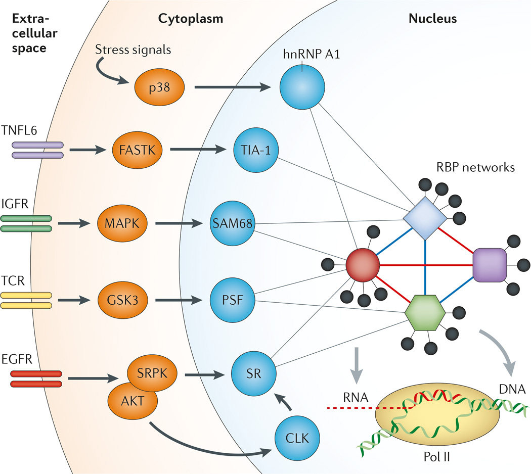
Sequence-specific RNA-binding proteins (RBPs) bind to pre-mRNA to control alternative splicing, but it is not yet possible to read the 'splicing code' that dictates splicing regulation on the basis of genome sequence. Each alternative splicing event is controlled by multiple RBPs, the combined action of which creates a distribution of alternatively spliced products in a given cell type. As each cell type expresses a distinct array of RBPs, the interpretation of regulatory information on a given RNA target is exceedingly dependent on the cell type. RBPs also control each other's functions at many levels, including by mutual modulation of their binding activities on specific regulatory RNA elements. In this Review, we describe some of the emerging rules that govern the highly context-dependent and combinatorial nature of alternative splicing regulation.


Figures

Figure 1
Figure 1. Regulation of alternative splicing by cis-acting enhancers and silencers
a | A pre-mRNA substrate has two 5′ splice sites (5′SSs) and two 3′SSs that are engaged in competition with each other for assembly of splicing complexes, which causes the alternative exon in the middle to be either included or skipped in the final mRNA products. In a given cell type, the ratio of included and skipped isoforms varies, and such variation is regulated by a large array of splicing regulators. b | Most exons in mammals, including both constitutive and alternative ones, are recognized by U1 small nuclear ribonucleoprotein (snRNP) binding at the 5′SS and U2 snRNP binding at the 3′SS that enhance one another. This exon definition process determines the selection of functional splice sites in the genome, which is subjected to modulation by various RNA-binding splicing regulators. c | Upon initial splice site selection, functional splice sites are paired to allow subsequent steps of spliceosome assembly to occur. Such a pairing process can also be regulated to generate alternatively spliced mRNA isoforms. d | Cis-acting RNA elements that positively or negatively influence splice site selection are shown. Depending on their locations and functions, they are referred to as exonic splicing enhancers (ESEs), exonic splicing silencers (ESSs), intronic splicing enhancers (ISEs) and intronic splicing silencers (ISSs). e | A selection of ESEs from a random sequence library are inserted into an alternative exon in a splicing reporter. In this setting, the middle exon is weakly included. From a random pool of sequences that have been individually inserted in the alternative exon, specific ESEs can be isolated, amplified and reselected from the mRNA product pool generated by in vitro splicing or from transfected cells that contain the alternative exon. f–h | Selection of ESSs (part f), ISEs (part g) and ISSs (part h) are shown. In each case, a sequence from a random sequence library is inserted into different locations in a splicing reporter that splits the GFP gene (green) into two halves. Upon transfection into an experimental cell line, GFP-positive cells are selected, and the pool of inserted sequences are then amplified by PCR and sequenced to identify sequence motifs that can modulate splicing.
Figure 2
Figure 2. Regulated splicing through controlling the assembly of the core splicing machinery
All splicing regulators must exert their activities, either directly or indirectly, through the core splicing machinery. a | Modulation of small nuclear ribonucleoprotein (snRNP) assembly can change the pool of active splicing components in the cell, thereby influencing alternative splice sites that are particularly sensitive to the levels of spliceosome components. b | The available functional splice sites in different pre-mRNAs — including the branchpoint sequence (BPS), polypyrimidine tract (PPT) and AG sequence in the 3′ splice site (3′SS), and the GU sequence in the 5′SS — are all competing for the splicing machinery. As a result, strong splice sites may titrate core components of the splicing machinery away from weak splice sites, thus indirectly influencing global splicing patterns in the cell. c | After initial recognition of splicing signals, the functional sites have to be efficiently paired for next steps of spliceosome assembly to take place, which also represents key steps for regulation. The binding of polypyrimidine-tract binding protein (PTB) to an intronic splicing silencer (ISS) inhibits intron definition. d | The U1 snRNP is about tenfold higher in abundance than other splicing snRNPs in mammalian cells. This may allow efficient recognition of most 5′SSs in both constitutive and alternative exons, as well as protection of inappropriate polyadenylation sites. However, the U1 snRNP has to be released later in the spliceosome cycle, which is essential for the formation of the catalytic core in the spliceosome. Interference of U1 release by heterogeneous nuclear ribonucleoprotein L (hnRNP L) and hnRNP A1 can thus also inhibit the selection of a regulated splice site, which acts as a repressive complex. ESE, exonic splicing enhancer; ESS, exonic splicing silencer; SF3A, splicing factor 3A; U2AF65, U2 auxiliary factor 65 kDa subunit.
Figure 3
Figure 3. Rules for context-dependent and position-sensitive regulation of alternative splicing
a | In general, the alternative exon (in the middle) is associated with weak splice site signals, which render partial selection by core components of the splicing machinery. A small U2 small nuclear ribonucleoprotein (snRNP) is illustrated to show its inefficient interaction with the alternative splice site. The flanking sites, which are strongly recognized by the splicing machinery, are engaging in competition with the alternative splice sites. A large U2 snRNP is illustrated to show its strong interaction with the flanking 3′ splice site (3′SS). According to the threshold rule, a partially compromised splicing machinery would selectively affect the alternative splice sites relative to the competing sites, thus leading to induced exon skipping. b | Exon-bound SR proteins and heterogeneous nuclear ribonucleoproteins (hnRNPs) have opposite effects on the recognition of the adjacent splice sites by the core splicing machinery. c | For many sequence-specific splicing factors (SFs) — for example, NOVA — their binding on exons generally inhibits the selection of the alternative exon. They are inhibitory when they are bound to an upstream intronic region but enhance the selection of the alternative exon when bound to a downstream intronic region. The mechanism for the polarity effect remains to be fully understood. d | Enhanced recognition of the alternative splice site induces exon inclusion, whereas increased recognition of the flanking competing splice site causes exon skipping. ESE, exonic splicing enhancer; ESS, exonic splicing silencer.
Figure 4
Figure 4. Cooperation and competition of splicing factors in regulated splicing
a | Distinct splicing regulators may bind to adjacent splicing regulatory elements (SREs) in a cooperative manner, which may be mediated, at least partly, by their protein–protein interactions (indicated by two-headed arrows). b | The same cis-acting SREs, regardless of whether they are located in exons or introns, may be recognized by related RNA-binding splicing regulators. This results in competition between related splicing regulators. c | An increasing amount of evidence suggests that the speed of transcription elongation may influence splice site selection, which may create window of opportunities for both positive and negative splicing regulators to recognize their binding sites co transcriptionally, thus modulating alternative splicing during these integrated events in gene expression. 3′SS, 3′ splice site; ESE, exonic splicing enhancer; ESS, exonic splicing silencer; hnRNP, heterogeneous nuclear ribonucleoprotein; ISS, intronic splicing silencer; Pol II, RNA polymerase II.
Figure 5
Figure 5. Signal transduction to regulate alternative splicing in the nucleus
Both internal and external signals can influence alternative splicing through post-transcriptional modification of specific splicing regulators as illustrated, which may modulate both protein–protein and protein–RNA interactions to produce a network response to affect many regulated splicing events. In terms of signal transduction pathways, the SR protein system seems to have a dedicated pathway through SR protein kinases (SRPKs) and CDC-like kinases (CLKs) in the cytoplasm and the nucleus, respectively. These kinases are responsible for controlling the phosphorylation state of SR proteins to modulate their activities in splicing. Various phosphatases may be also involved to antagonize the activity of these splicing kinases. EGFR, epidermal growth factor receptor; FASTK, Fas-activated serine/threonine kinase; GSK3, glycogen synthase kinase 3; hnRNP A1, heterogeneous nuclear ribonucleoprotein A1; IGFR, insulin-like growth factor receptor; MAPK, mitogen-activated protein kinase; p38 is a MAPK involved in the DNA damage response; Pol II, RNA polymerase II; PSF, PTB (polypyrimidine-tract binding protein)-associated splicing factor; RBP, RNA-binding protein; TCR, T cell receptor; TIA 1, nucleolysin TIA 1 isoform p40; TNFL6, tumour necrosis factor ligand superfamily member 6 (also known as FasL).

References

    1. Penny D, Hoeppner MP, Poole AM, Jeffares DC. An overview of the introns-first theory. J. Mol. Evol. 2009;69:527–540. - PubMed
    1. Ast G. How did alternative splicing evolve? Nature Rev. Genet. 2004;5:773–782. - PubMed
    1. Keren H, Lev-Maor G, Ast G. Alternative splicing and evolution: diversification, exon definition and function. Nature Rev. Genet. 2010;11:345–355. - PubMed
    1. Brown JB, et al. Diversity and dynamics of the Drosophila transcriptome. Nature. 2014 http://dx.doi. org/10.1038/nature12962. - DOI - PMC - PubMed
    1. Barbosa-Morais NL, et al. The evolutionary landscape of alternative splicing in vertebrate species. Science. 2012;338:1587–1593. - PubMed

Publication types

Substances