. 2013 Nov 21:3737:473-87.
doi: 10.11646/zootaxa.3737.4.8.

Redescription of Alatina alata (Reynaud, 1830) (Cnidaria: Cubozoa) from Bonaire, Dutch Caribbean

Affiliations

Redescription of Alatina alata (Reynaud, 1830) (Cnidaria: Cubozoa) from Bonaire, Dutch Caribbean

Cheryl Lewis et al. Zootaxa. .

Abstract

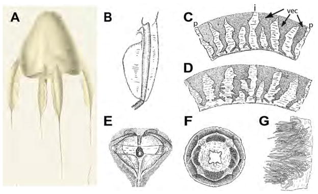
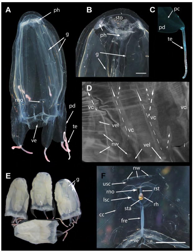
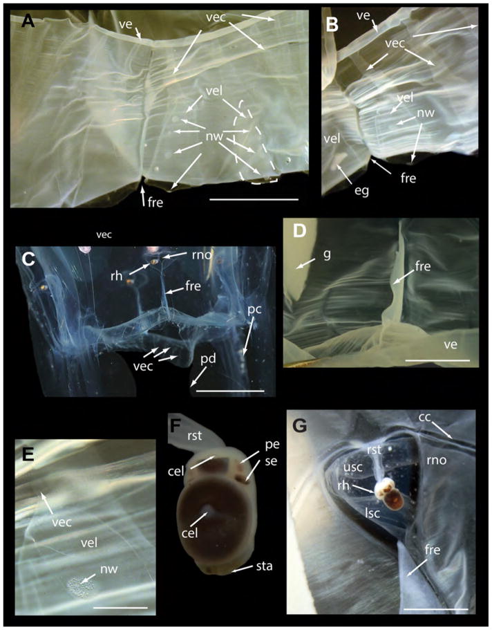
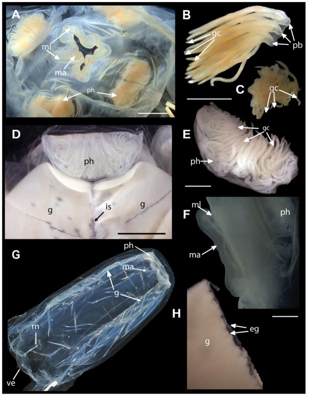
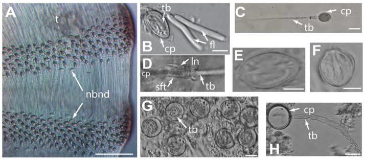
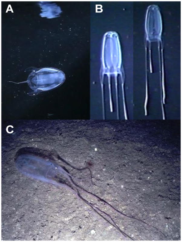
Here we establish a neotype for Alatina alata (Reynaud, 1830) from the Dutch Caribbean island of Bonaire. The species was originally described one hundred and eighty three years ago as Carybdea alata in La Centurie Zoologique-a monograph published by René Primevère Lesson during the age of worldwide scientific exploration. While monitoring monthly reproductive swarms of A. alata medusae in Bonaire, we documented the ecology and sexual reproduction of this cubozoan species. Examination of forty six A. alata specimens and additional archived multimedia material in the collections of the National Museum of Natural History, Washington, DC revealed that A. alata is found at depths ranging from surface waters to 675 m. Additional studies have reported it at depths of up to 1607 m in the tropical and subtropical Atlantic Ocean. Herein, we resolve the taxonomic confusion long associated with A. alata due to a lack of detail in the original description and conflicting statements in the scientific literature. A new cubozoan character, the velarial lappet, is described for this taxon. The complete description provided here serves to stabilize the taxonomy of the second oldest box jellyfish species, and provide a thorough redescription of the species.


Figures

FIGURE 1
FIGURE 1
Line drawings of A. alata reproduced from Reynaud (1830) and Bigelow (1938) (as Carybdea alata). A. Whole body showing bell, tentacles and pedalia (reproduced from Reynaud (1830). B–G, Reproduced from Bigelow (1938). B. Pedalium, C. Velarial canals (BH=70 mm), D. Velarial canals (BH=90 mm), E. Rhopalial niche with T–shaped opening, F. Bell apex showing crescentric gastric phacellae, G. Dissected gastric phacella (comprising gastric cirri). Abbreviations: i=interradius (location of pedalium), p=perradius (location of frenulum), vec=velarial canal (three per quadrant, simple to branching).
FIGURE 2
FIGURE 2
A. alata neotype (USNM 1195802 unless otherwise specified) from Kralendijk, Bonaire, The Netherlands. A. Mature female medusa (live), whole body (live, BW=40, BH=70). B. Apical portion showing stomach and crescentric gastric phacellae, visible in each corner, and wide central manubrium opening into subumbrella in live medusa. C. Pedalium and tentacle of live medusa. D. Velarial canals (3 per octant) in preserved medusa: each bears a row of 3 nematocyst warts on proximal first half to two thirds (on the velarial lappets) (dashed arrows indicate the main three velarial canals and branches extending from the main base of each canal). E. USNM 1195803–1195806, preserved mature medusae from the same locality as the neotype. F. T–shaped rhopalial niche opening with nematocyst warts on upper and lower scale coverings in live medusa. Live photographs by T. Peters (Fish Eye Photography, Bonaire) & A. Yanagihara. Abbreviations: cc=circular canal, fre=frenulum, g=gonads, i=interradius, lsc=lower scale of rhopalial niche covering, nw=nematocyst wart, ph=gastric phacellae (comprises gastric cirri), pc=pedalial canal, pd=pedalium, rh=rhopalium, rno=rhopalial niche opening, rst=rhopalial stalk, sta=statocyst, sto=stomach, te=tentacle, usc=upper scale of rhopalial niche covering. Scale bars: 5 mm (B & F).
FIGURE 3
FIGURE 3
Alatina alata from Kralendijk, Bonaire, The Netherlands (same location as neotype). A. & B. USNM 1195807, velarial canals (3 per octant) in preserved medusa, simple to branching (one outlined with dotted line in Fig 3A). Each canal bears a row of 3 nematocyst warts on the proximal first half to two thirds, i.e., on the velarial lappets. A clump of disassociated eggs is seen within the velarial canal on the left. C. USNM 1195802 (neotype), velarium of live medusa, partially pushed out revealing velarial canals. D. USNM 1195806, double layered frenulum anchoring the subumbrella to velarium at perradius. E. USNM 1195807, nematocyst wart on velarial lappet filled with isorhiza nematocysts. F. USNM 1195804, rhopalium dissected from preserved medusa, bears two median complex eyes, two upper lateral pit eyes, and two lower lateral slit eyes. G. USNM 1195806, subumbrella view of rhopalial window of preserved medusa, outline of rhopalial niche opening visible. Live photographs by T. Peters (Fish Eye Photography, Bonaire) & A. Yanagihara. Abbreviations: cel=complex eye lens, fre=frenulum, g=gonads, nw=nematocyst wart, pc=pedalial canal, pe=pit eye, pd=pedalium, rh=rhopalium, rno=rhopalial niche opening, rst=rhopalial stalk, se=slit eye, sta=statocyst, usc=upper scale of rhopalial niche covering. ve=velarium, vec=velarial canal, vel=velarial lappet. Scale bars: 5 mm (A & D), 10 mm (C), 1 mm (E), 3 mm (G).
FIGURE 4
FIGURE 4
Alatina alata specimens, preserved and live (all except A–C from Kralendijk, Bonaire (The Netherlands) the same locality as the neotype). A. USNM 53659, oral view of manubrium and four crescentric gastric phacellae in each corner of the stomach (BW=19 mm, BH=47 mm). B. USNM 53659, dissected tuft of gastric cirri from crescentric gastric phacella. C. USNM 53659, dissected gastric cirri, appearing as central mass in stomach of developing medusa (BW=10 mm, BH=22 mm). D. USNM 1195807, gastric phacellae and adjacent ripe paired gonads in preserved female medusa. E. USNM 1195807, dissected crescentric gastric phacella from mature medusa. F. USNM 1195806, lateral view of manubrium with mouth lips curled up. G. Live mature medusa, gonads beginning to rupture following a reproductive swarming episode (live, BW=60, BH=90). H. USNM 1195807, gonads dissected from mature female, bilayer of mature oocytes visible along adradial point of rupture. Live photographs by T. Peters (Fish Eye Photography Bonaire) and A. Yanagihara. (Fish Eye Photography, Bonaire). Abbreviations: eg=eggs, gc=gastric cirri, ma=manubrium, ml=mouth lips, pb=primary branch, ph=gastric phacellae (comprises gastric cirri), is=interradial septum. Scale bars: 5 mm (A & D), 2mm (B, C, E, F, H).
FIGURE 5
FIGURE 5
Cnidome of Alatina alata (all extracted from preserved material unless specified otherwise). A. Nematocyst bands along the length of the tentacle of a live specimen collected in Bonaire (June 2013). B. & E. Undischarged, microbasic heterotrichus p–euryteles extracted from tentacles; filaments associated with euryteles in B (USNM 1195807). C. Discharged microbasic heterotrichus p–eurytele extracted from tentacles of live medusa. D. Lancet contiguous with discharged tubule of microbasic heterotrichus p–eurytele extracted from tentacles (USNM 1195805) F. Small undischarged birhopaloid found in tentacles of preserved specimen USNM 1195807. G. Large undischarged holotrichus isorhizas found in nematocysts warts (i.e., bell warts & velarial lappet warts (USNM 1195802, neotype). H. Large discharged holotrichus isorhizas found in bell warts (USNM 1195802). H. Abbreviations: cp=capsule, fl=filaments, ln=lancet, nbnd=nematocyst band, sft=shaft, tb=tubule. Scale bars: ~150 μm (A), 10 μm (B, E, F), 25 μm (C), 20 μm (G, H).
FIGURE 6
FIGURE 6
Alatina alata in situ images. A. A. alata at a depth of 2 m from Kralendijk, Bonaire, The Netherlands. Reflection on the underside of the surface of the water seen above the medusa (frame grab from video by D. Karamehmedovic & A. Yanagihara). B. Series of digital frame grabs taken from video footage of a mature A. alata medusa actively ascending in the water column; filmed at a depth of 500–540 m, West off Gorda Cay, Bahamas, from Johnson Sea Link I manned submersible (video voucher USNM 1195809). B. Photograph of A. alata medusa oriented parallel to the ocean bottom, photographed by the Remotely Operated Vehicle SeaROVER at a depth of about 100 m in the Gulf of Mexico (USNM 1005621). No specimen was collected as a museum voucher for multimedia material.

References

    1. Agassiz L. Contributions to the natural history of the United States of America. IV. Little Brown; Boston: 1862. [accessed 2 Oct 2013]. p. 380.p. 19. Available from: http://www.biodiversitylibrary.org/item/54510#page/9/mode/1up.
    1. Arneson AC. MS Thesis. University of Puerto Rico, Mayaguez, Commonwealth of Puerto Rico; USA: 1976. Life history of Carybdea alata Reynaud, 1830 (Cubomedusae) p. 43.
    1. Arneson AC, Cutress CE. Life history of Carybdea alata Reynaud, 1830 (Cubomedusae) In: Mackie GO, editor. Coelenterate Ecology and Behavior. Plenum Press; New York, NY: 1976. pp. 227–236.
    1. Bauchot ML, Daget J, Bauchot R. L’ichthyologie en France au début du XIXe siècle. L’Histoire naturelle des Poissons de Cuvier et Valenciennes. Bulletin du Muséum national d’Histoire naturelle, Paris, section A. 1990;(supplément 12):3–142. http://dx.doi.org/10.3366/anh.1993.20.1.139a. - DOI
    1. Bentlage B. Carybdea alata auct. (Cubozoa): rediscovery of the Alatina grandis type. Zootaxa. 2010;2713:52–54.

LinkOut - more resources