. 2014 Sep 26;289(39):27146-27158.
doi: 10.1074/jbc.M114.572172. Epub 2014 Aug 11.

Thrombin cleavage of osteopontin disrupts a pro-chemotactic sequence for dendritic cells, which is compensated by the release of its pro-chemotactic C-terminal fragment

Affiliations

Thrombin cleavage of osteopontin disrupts a pro-chemotactic sequence for dendritic cells, which is compensated by the release of its pro-chemotactic C-terminal fragment

Zhifei Shao et al. J Biol Chem. .

Abstract

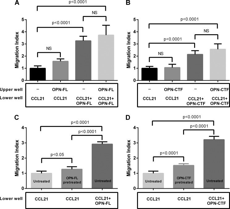
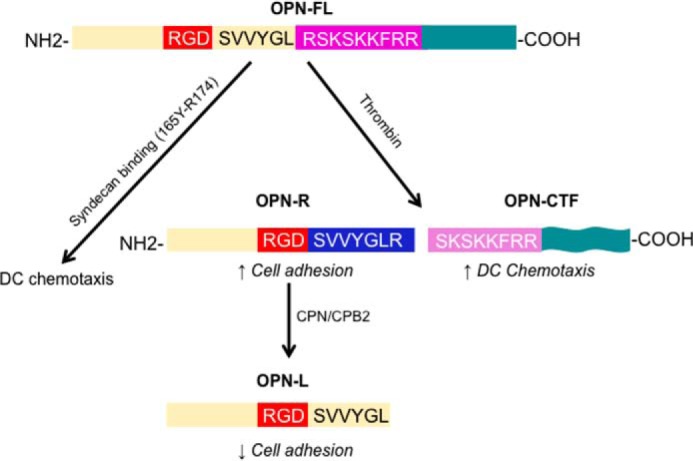
Thrombin cleavage alters the function of osteopontin (OPN) by exposing an integrin binding site and releasing a chemotactic C-terminal fragment. Here, we examined thrombin cleavage of OPN in the context of dendritic cell (DC) migration to define its functional domains. Full-length OPN (OPN-FL), thrombin-cleaved N-terminal fragment (OPN-R), thrombin- and carboxypeptidase B2-double-cleaved N-terminal fragment (OPN-L), and C-terminal fragment (OPN-CTF) did not have intrinsic chemotactic activity, but all potentiated CCL21-induced DC migration. OPN-FL possessed the highest potency, whereas OPNRAA-FL had substantially less activity, indicating the importance of RGD. We identified a conserved (168)RSKSKKFRR(176) sequence on OPN-FL that spans the thrombin cleavage site, and it demonstrated potent pro-chemotactic effects on CCL21-induced DC migration. OPN-FLR168A had reduced activity, and the double mutant OPNRAA-FLR168A had even lower activity, indicating that these functional domains accounted for most of the pro-chemotactic activity of OPN-FL. OPN-CTF also possessed substantial pro-chemotactic activity, which was fully expressed upon thrombin cleavage and its release from the intact protein, because OPN-CTF was substantially more active than OPNRAA-FLR168A containing the OPN-CTF sequence within the intact protein. OPN-R and OPN-L possessed similar potency, indicating that the newly exposed C-terminal SVVYGLR sequence in OPN-R was not involved in the pro-chemotactic effect. OPN-FL and OPN-CTF did not directly bind to the CD44 standard form or CD44v6. In conclusion, thrombin cleavage of OPN disrupts a pro-chemotactic sequence in intact OPN, and its loss of pro-chemotactic activity is compensated by the release of OPN-CTF, which assumes a new conformation and possesses substantial activity in enhancing chemokine-induced migration of DCs.

Keywords: Chemokine; Dendritic Cell; Migration; Osteopontin; Thrombin.


Figures

FIGURE 1.
FIGURE 1.
Schematic structure and purity of recombinant human osteopontins. A, schematic of recombinant mature full-length human OPN (amino acids 1–314) (OPN-FL), RGD-mutated OPN-FL (OPNRAA-FL), mature full-length human OPN mutant in which Arg168 was substituted with Ala (OPN-FLR168A), and double mutant OPNRAA-FLR168A and thrombin-cleaved OPN N-terminal fragment 1–168 (OPN-R), thrombin- and CPB2-cleaved OPN N-terminal fragment 1–167 (OPN-L), thrombin-cleaved OPN C-terminal fragment 169–314 (OPN-CTF) (all the above recombinant proteins have GST fused to their N termini), and thrombin-cleaved OPN C-terminal fragment with free N terminus and His6 tag at its C terminus (OPN-CTFHis6). B, recombinant osteopontin proteins were purified by affinity chromatography using a glutathione-Sepharose column. A total of 2 μg of purified proteins were separated by SDS-PAGE and stained by Coomassie Brilliant Blue.
FIGURE 2.
FIGURE 2.
Cell surface phenotype of human monocyte-derived DCs. Flow cytometry analysis of DC surface expression of CD44, CD44v6, αv, αvβ3, αvβ5, β1, α4β1, and α9β1 integrins, CD11c, HLA-DR, CD1a, and CCR7 on LPS-stimulated mature human monocyte-derived DCs. Gray-filled histograms, isotype controls; black empty histograms, target antigens.
FIGURE 3.
FIGURE 3.
OPN-FL and thrombin-cleaved N-terminal fragments enhance DC migration toward lymphatic chemokines CCL21 and CCL19. DCs were added to the upper chamber of a Transwell plate. CCL21 (50 ng/ml or 4.2 nm) and/or OPN (100 nm) was added into serum-free culture medium in the lower chamber to create a concentration gradient. A, DC migration to OPN or thrombin-cleaved OPN fragments was compared with migration of DCs to CCL21 (n = 9–24; data pooled from seven independent experiments; *, p < 0.0001, CCL21 versus each OPN). B, DC migration to CCL21 in the presence of various concentrations of OPN-FL (n = 3–6). The level of migration observed in CCL21 alone was subtracted for curve fitting. C, DC migration to 50 ng/ml CCL21 in the presence of 100 nm OPN was compared with migration to CCL21 alone (n = 6–20; data pooled from six independent experiments). D, DC migration to 25 ng/ml CCL19 in the presence of 100 nm OPN was compared with migration to CCL19 alone (n = 6; data are representative of three independent experiments). In all experiments, the value from medium alone was subtracted as background. Migrated cells in the lower chamber of the Transwell were normalized relative to cells migrating to CCL21 or CCL19. Data are shown as mean with CI (error bars).
FIGURE 4.
FIGURE 4.
DC migration toward CCL21 in the presence of thrombin-cleaved OPN C-terminal fragment and its overlapping peptides. A, DC migration to CCL21 (50 ng/ml or 4.2 nm) in the presence of 100 nm OPN-CTF, OPN-FL, OPN-R, or a combination of 100 nm OPN-CTF + 100 nm OPN-R (n = 6–25; data pooled from three independent experiments). B, DC migration to CCL21 (50 ng/ml or 4.2 nm) in the presence of various concentrations of OPN-CTF (n = 3–4). The level of migration observed in CCL21 alone was subtracted. C, DC migrations to various concentrations (0.3–200 nm) of CCL21 in the presence of 100 nm OPN-CTF (n = 4). D, DC migration to CCL21 in the presence of 2 μm overlapping peptides P0–P7 derived from OPN-CTF. (See Table 1 for peptide sequences; n = 6–17; data pooled from three independent experiments). Data are shown as mean with CI (error bars). *, p < 0.05; **, p < 0.01; ****, p < 0.0001.
FIGURE 5.
FIGURE 5.
Sequence spanning thrombin cleavage site is pro-chemotactic. A, DC migration to CCL21 (50 ng/ml or 4.2 nm) in the presence of 100 nm OPN-FL, OPNRAA-FL, OPN-FLR168A, and OPNRAA-FLR168A (n = 4–20; data pooled from four independent experiments). B, DC migration to 50 ng/ml CCL21 was measured in the presence of various concentrations of P0, P0R168A, P0K170A, P0K172A/K173A, and P0reversed (see Table 1 for peptide sequences; n = 10; data are representative of three independent experiments). C, DC migration to 50 ng/ml CCL21 was measured in the presence of various concentrations of P0, PR168-T185, PS169-T185, and PS168-T185-scrambled (see Table 1 for peptide sequences; n = 9–10; data are representative of two independent experiments). Data are shown as mean with CI (error bars).
FIGURE 6.
FIGURE 6.
Thrombin cleavage and C-terminal modification altered pro-chemotactic activity of OPN-CTF. A, DC migrated to CCL21 (50 ng/ml or 4.2 nm) in the presence of various concentrations of OPN-CTF and OPNRAA-FLR168A (n = 4–8; data pooled from two independent experiments); B, DC migration to CCL21 (50 ng/ml or 4.2 nm) in the presence of various concentrations of OPN-CTF and OPN-CTFHis6 (n = 8; data pooled from two independent experiments). Data are shown as mean with CI (error bars). C, representative surface plasmon resonance sensorgrams of OPN-FL, OPN-CTF, OPNRAA-FLR168A, and OPN-CTFHis6 binding to immobilized MAB197P. Langmuir fits to the kinetic data are shown in Table 2. Response (resonance units (RU)) is plotted against time. Injected OPN concentrations (from top to bottom traces) are 1000, 300, 100, 30, and 10 nm.
FIGURE 7.
FIGURE 7.
Maximum effect of OPN-FL and OPN-CTF requires temporal and physical co-existence of OPN and CCL21. DC migration to CCL21 (50 ng/ml or 4.2 nm) in the presence of 100 nm OPN-FL (A) or 100 nm OPN-CTF (B) in the upper well, lower well, or both upper and lower wells (n = 16; data pooled from two independent experiments). DCs were treated with 100 nm OPN-FL (C) or 100 nm OPN-CTF (D) for 1 h, followed by washing with PBS to remove residual OPN-FL or OPN-CTF. OPN-pretreated and untreated DCs were loaded onto upper wells of Transwell plates, in which the lower wells contained CCL21 (50 ng/ml) or CCL21 (50 ng/ml) and 100 nm OPN-FL/OPN-CTF (n = 8–22; data pooled from three independent experiments). Error bars, CI.
FIGURE 8.
FIGURE 8.
OPN-FL or OPN-CTF does not alter the amplitude of CCL21-induced calcium transients. Mature DCs were loaded with the calcium indicator Fluo-8 and plated onto a 96-well plate. A, various concentrations (20 pm to 200 nm) of CCL21 were added to DC suspensions to demonstrate a dose-dependent response of CCL21-induced calcium transients (EC50 = 3.56 nm; n = 3–4 at each concentration). B, CCL21 (50 ng/ml or 4.1 nm) was added into DC suspension in which OPN-FL or OPN-CTF was present (n = 11). The green fluorescence (excitation, 485 nm; emission, 488 nm) was recorded before and after the addition of CCL21. ΔE488 indicates the maximum change in fluorescence upon CCL21 stimulation. Error bars, CI.
FIGURE 9.
FIGURE 9.
Anti-CD44 and CD44v6 antibodies blocked 50 ng/ml CCL21-induced migration in the absence of OPN. The cells were treated with 10 μg/ml of antibody for 15 min before adding them to the upper chamber. Antibody (10 μg/ml) was added to the lower chamber of the Transwell plate (n = 8–14; data pooled from two independent experiments). Data are shown as mean with CI (error bars). *, p < 0.05; ****, p < 0.0001.
FIGURE 10.
FIGURE 10.
Mouse bone marrow-derived DCs migrate to chemokines in the presence of OPN. A, WT mature BMDC migration toward CCL21 (100 ng/ml or 8.4 nm) supplemented with different OPN forms was compared with migration to CCL21 alone (n = 8). B, WT immature BMDC migration to CCL3 (50 ng/ml or 6.4 nm) supplemented with different OPN forms was compared with migration to CCL3 alone (n = 3). C, mature OPN−/− BMDC migration to 100 ng/ml CCL21 supplemented with different OPN forms was compared with migration to CCL21 alone (n = 3). Data are shown as mean with CI (error bars). *, p < 0.05; **, p < 0.01; ***, p < 0.001; ****, p < 0.0001.
FIGURE 11.
FIGURE 11.
Thrombin cleavage of OPN modulates chemokine-induced DC migration. At the initiation of inflammation, while DC is being activated, cell matrix proteoglycan-bound OPN will maintain its full-length form and exert its maximal potentiating effect on chemokine-induced DC migration, mediated by the 159RGD161 and 168RSKSKFRR176 sequences, despite the presence of thrombin. With further progression of inflammation and local generation of thrombin and CPB2, thrombin cleavage of OPN occurs, which will increase cell adhesion by exposing SVVYGLR and allowing better access to RGD in OPN-R while reducing its potentiating effect on DC migration. Further cleavage of OPN-R by CPB2 (or CPN) inactivates SVVYGLR, converting it to OPN-L, and thus decreases cell adhesion mediated by α4β1 and α9β1 integrins. While thrombin cleavage disrupts the RSKSKKFRR pro-chemotactic domain in intact OPN, it releases OPN-CTF (Ser169–Asn314), which assumes a new conformation and possesses substantial pro-chemotactic activity, compensating for the loss of the pro-chemotactic activity in OPN-R and OPN-L.

Similar articles

Cited by

References

    1. Lund S. A., Giachelli C. M., Scatena M. (2009) The role of osteopontin in inflammatory processes. J. Cell Commun. Signal. 3, 311–322 - PMC - PubMed
    1. Sharif S. A., Du X., Myles T., Song J. J., Price E., Lee D. M., Goodman S. B., Nagashima M., Morser J., Robinson W. H., Leung L. L. (2009) Thrombin-activatable carboxypeptidase B cleavage of osteopontin regulates neutrophil survival and synoviocyte binding in rheumatoid arthritis. Arthritis Rheum. 60, 2902–2912 - PMC - PubMed
    1. Weiss J. M., Renkl A. C., Maier C. S., Kimmig M., Liaw L., Ahrens T., Kon S., Maeda M., Hotta H., Uede T., Simon J. C. (2001) Osteopontin is involved in the initiation of cutaneous contact hypersensitivity by inducing Langerhans and dendritic cell migration to lymph nodes. J. Exp. Med. 194, 1219–1229 - PMC - PubMed
    1. Grassinger J., Haylock D. N., Storan M. J., Haines G. O., Williams B., Whitty G. A., Vinson A. R., Be C. L., Li S., Sørensen E. S., Tam P. P., Denhardt D. T., Sheppard D., Choong P. F., Nilsson S. K. (2009) Thrombin-cleaved osteopontin regulates hemopoietic stem and progenitor cell functions through interactions with α9β1 and α4β1 integrins. Blood 114, 49–59 - PubMed
    1. Bruemmer D., Collins A. R., Noh G., Wang W., Territo M., Arias-Magallona S., Fishbein M. C., Blaschke F., Kintscher U., Graf K., Law R. E., Hsueh W. A. (2003) Angiotensin II-accelerated atherosclerosis and aneurysm formation is attenuated in osteopontin-deficient mice. J. Clin. Invest. 112, 1318–1331 - PMC - PubMed

Publication types