. 2014 Sep 26;289(39):27134-27145.
doi: 10.1074/jbc.M114.594812. Epub 2014 Aug 11.

p53 is required for cisplatin-induced processing of the mitochondrial fusion protein L-Opa1 that is mediated by the mitochondrial metallopeptidase Oma1 in gynecologic cancers

Affiliations

p53 is required for cisplatin-induced processing of the mitochondrial fusion protein L-Opa1 that is mediated by the mitochondrial metallopeptidase Oma1 in gynecologic cancers

Bao Kong et al. J Biol Chem. .

Abstract

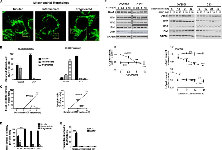
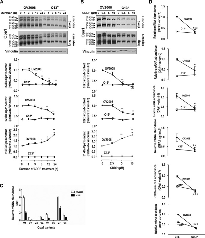
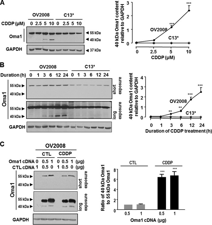
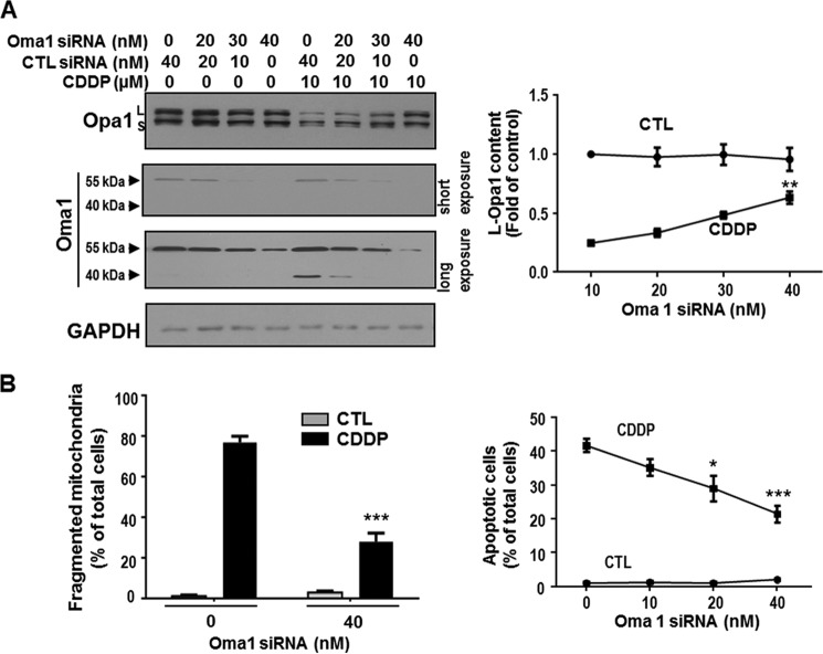
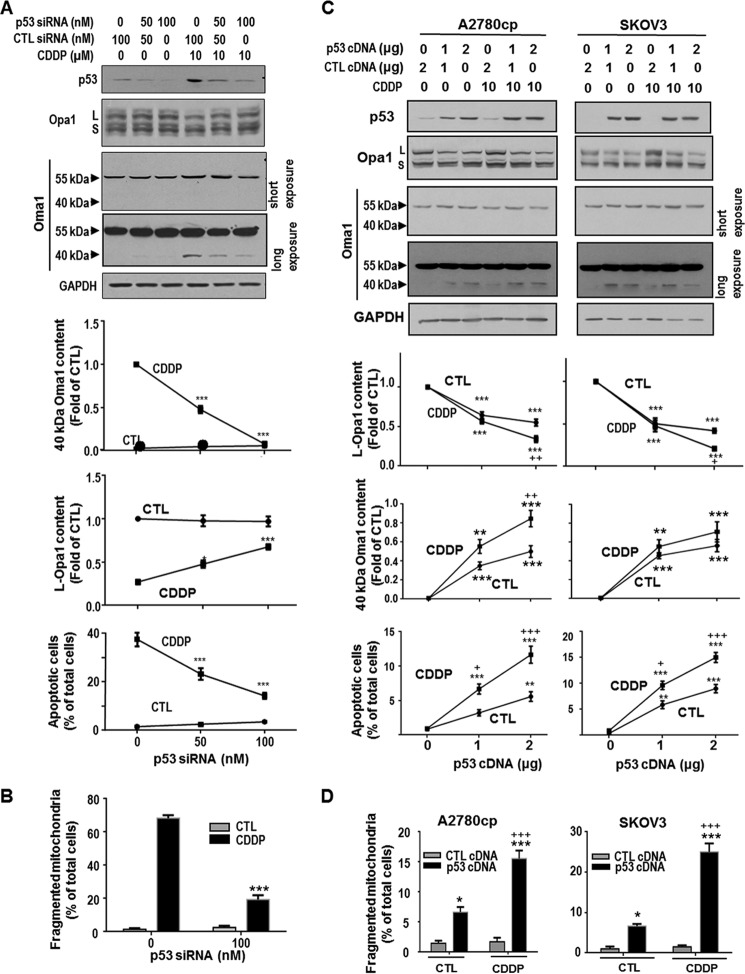
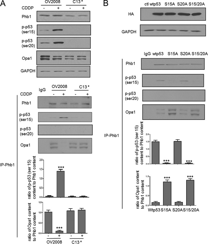
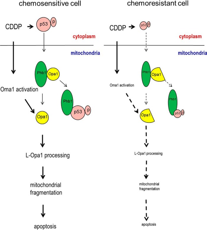
Mitochondria are highly dynamic organelles, and mitochondrial fission is a crucial step of apoptosis. Although Oma1 is believed to be responsible for long form Opa1 (L-Opa1) processing during mitochondrial fragmentation, whether and how Oma1 is involved in L-Opa1 processing and participates in the regulation of chemoresistance is unknown. Chemosensitive and chemoresistant ovarian (OVCA) and cervical (CECA) cancer cells were treated with cisplatin (CDDP). Mitochondrial dynamics and protein contents were assessed by immunofluorescence and Western blot, respectively. The requirements of Oma1 and p53 for CDDP-induced L-Opa1 processing, mitochondrial fragmentation, and apoptosis were examined by siRNA or cDNA. CDDP induces L-Opa1 processing and mitochondrial fragmentation in chemosensitive but not in chemoresistant cells. CDDP induced Oma1 40-kDa form increases in OV2008 cells, not in C13* cells. Oma1 knockdown inhibited L-Opa1 processing, mitochondrial fragmentation, and apoptosis. Silencing p53 expression attenuated the effects of CDDP in Oma1 (40 kDa) increase, L-Opa1 processing, mitochondrial fragmentation, and apoptosis in chemosensitive OVCA cells, whereas reconstitution of p53 in p53 mutant or null chemoresistant OVCA cells induced Oma1 (40 kDa) increase, L-Opa1 processing, mitochondrial fragmentation, and apoptosis irrespective of the presence of CDDP. Prohibitin 1 (Phb1) dissociates from Opa1-Phb1 complex and binds phosphorylated p53 (serine 15) in response to CDDP in chemosensitive but not chemoresistant CECA cells. These findings demonstrate that (a) p53 and Oma1 mediate L-Opa1 processing, (b) mitochondrial fragmentation is involved in CDDP-induced apoptosis in OVCA and CECA cells, and (c) dysregulated mitochondrial dynamics may in part be involved in the pathophysiology of CDDP resistance.

Keywords: Apoptosis; CDDP; Chemoresistance; Mitochondrial Fragmentation; Oma1; Opa1; Ovarian Cancer; Protein-Protein Interaction; p53.


Figures

FIGURE 1.
FIGURE 1.
Mitochondrial dynamics in chemosensitive and chemoresistant gynecologic cancer cells. A, mitochondrial phenotypes (tubular, intermediate, and fragmented) in CECA cells (OV2008 and C13*). Mitochondria were immunostained with Tom20 antibody. Mitochondrial phenotypes were examined by immunofluorescence confocal microscopy. The inset is an enlarged image of the boxed area. B, comparison of mitochondrial phenotypes in chemosensitive and chemoresistant CECA cells treated with CDDP. OV2008 and C13* cells were cultured with and without CDDP (10 μm, 0–6 h). Percentage of cells with tubular mitochondria in chemoresistant cells (C13*) is higher than in chemosensitive cells (OV2008) at 0 h. More than 100 cells were assessed in each experimental group (n = 3, ***, p < 0.001). C, comparison of mitochondrial fragmentation phenotype and apoptosis in chemosensitive and chemoresistant CECA cells treated with CDDP. OV2008 and C13* cells were cultured with CDDP (10 μm, 0–6 h). Mitochondrial phenotypes were examined by immunofluorescence confocal microscopy. OV2008 cells exhibited much higher level of fragmented mitochondria than C13* cells with CDDP treatment (n = 3, **, p < 0.01 at 3 h and ***, p < 0.001 at 6 h). At least 100 cells were assessed for each time point in each replicate. Apoptosis was examined by Hoechst assay. OV2008 cells exhibited higher apoptosis than C13* cells when treated with CDDP (n = 3, ***, p < 0.001 at 24 and 48 h). More than 300 cells were assessed in each experimental group. D, comparison of mitochondrial phenotypes in different OVCA cell lines. Chemoresistant A2780cp cells had more tubular mitochondria than chemosensitive A2780s cells (n = 3, ***, p < 0.001). Most SKOV3 and HEY cells exhibited tubular mitochondria. Over 100 cells were assessed in each experimental group. E, CDDP-induced mitochondria fragmentation in chemosensitive but not chemoresistant OVCA cells. A2780s, A2780cp, SKOV3, and HEY cells were cultured in the presence of CDDP (10 μm, 6 h) or control (CTL; DMSO) only. Mitochondrial phenotypes were examined by immunofluorescence confocal microscopy. CDDP induced significant mitochondria fragmentation in A2780s (***, p < 0.001, n = 3) but not in A2780cp, SKOV3, and HEY cells. At least 100 cells were assessed at each time point in each replicate. F, influence of CDDP on contents of mitochondrial fission proteins (Drp1 and Fis1) and mitochondrial fusion proteins (Mfn1, Mfn2, and Opa1) in CECA cells. OV2008 and C13* cells were cultured with CDDP (left panel, 0–10 μm, 24 h; right panel, 0–10 μm, 0–48 h), Opa1, Mfn1, Mfn2, Drp1, and Fis1 contents were examined by Western blot. Opa1 content (long form) significantly decreased in a concentration- and time-dependent manner in chemosensitive (OV2008) but not in chemoresistant (C13*) cells when treated with CDDP. L and S represent long and short form of Opa1, respectively. Contents of Mfn1, Mfn2, Drp1, and Fis1 were not altered by CDDP treatment. Representative Western blots of four independent experiments are shown.
FIGURE 2.
FIGURE 2.
Different forms of Opa1 in CECA cells. Chemosensitive (OV2008) and chemoresistant (C13*) CECA cells were cultured with CDDP for different durations (A; 10 μm, 0–24 h) or at different concentrations (B; 0–10 μm, 24 h). Contents of different forms of Opa1 and vinculin (loading control) were examined by Western blot. Five forms of Opa1 (95, 92, 88, 84, and 81 kDa) were recognized in OV2008 cells, whereas only three forms (95, 92, and 84 kDa) were recognized in C13* cells. CDDP significantly decreased contents of L-Opa1 forms (95 and 92 kDa), whereas it significantly increased S-Opa1 content (81 kDa) in a time-dependent (A; *, p < 0.05; **, p < 0.01, n = 4) and concentration-dependent (B; *, p < 0.05; **, p < 0.01; ***, p < 0.001, n = 3) manner in OV2008 cells but not in C13* cells. C, OV2008 and C13* cells were cultured with CDDP (10 μm, 6 h; DMSO as control (CTL)), and the abundance of each Opa1 splice form was examined by quantitative PCR. Forms 1, 2, 4, 5, 7, and 8 were expressed in OV2008 and C13* cells, and the abundance of the above forms are higher in OV2008 cells than C13* cells (p < 0.05, n = 3). V with a number represents specific form of Opa1. D, CDDP decreased all Opa1 splice forms detected in OV2008 cells, not in C13* cells (p < 0.05, n = 3).
FIGURE 3.
FIGURE 3.
Different forms of Oma1 in chemosensitive and chemoresistant CECA cells. A and B, OV2008 and C13* cells were cultured with CDDP at different concentrations (A; 0–10 μm, 24 h) or for different duration (B; 0–24 h, 10 μm). Contents of Oma1 and GAPDH (loading control) were examined by Western blotting (n = 3). Two forms of Oma1 (55 and 40 kDa) were present in OV2008 cells but only one (55 kDa) in C13* cells when cultured in the presence of CDDP. Oma1 contents (40 kDa) in OV2008 cells but not in C13* cells significantly increased in the presence of CDDP in a concentration- and time-dependent manner. C, OV2008 cells were transfected with Myc-tagged Oma1 cDNA or control cDNA (CTL; vector) and subsequently cultured with CDDP (10 μm, 24 h) or control (DMSO). Two Myc bands (55 and 40 kDa) were detected by Western blot with anti-Myc antibody, with the ratio of 40-kDa band to 55-kDa band significantly increased in the presence of CDDP (***, p < 0.001, n = 3).
FIGURE 4.
FIGURE 4.
Oma1 knockdown attenuated CDDP-induced loss in L-Opa1 content, mitochondrial fragmentation, and apoptosis in OV2008 cells. A, OV2008 cells were treated with Oma1 siRNA or control siRNA (CTL; scramble siRNA) (0–40 nm, 24 h) and cultured with CDDP (10 μm, 24 h) or control (DMSO). L-Opa1 and Oma1 contents were examined by Western blot. Oma1 siRNA (40 nm) significantly attenuated the loss of L-Opa1 content induced by CDDP. B, left: OV2008 cells were treated with Oma1 siRNA or control siRNA (scramble siRNA) (0–40 nm, 24 h) and subsequently with CDDP (10 μm, 6 h) or control (DMSO). Mitochondria were immunostained with anti-Tom20 antibody and examined by immunofluorescence confocal microscopy. Oma1 siRNA significantly decreased mitochondrial fragmentation induced by CDDP (***, p < 0.001. n = 3). More than 100 cells were assessed in each experimental group. Right: OV2008 cells were treated with Oma1 siRNA or control siRNA (scramble siRNA) (40 nm, 24 h) and subsequently with CDDP (10 μm, 24 h) or control (DMSO). Apoptosis was examined by Hoechst assay. Oma1 siRNA significantly decreased apoptosis induced by CDDP (***, p < 0.001; n = 3). More than 300 cells were assessed in each experimental group.
FIGURE 5.
FIGURE 5.
p53-dependent action of CDDP on Oma1 (40 kDa) and L-Opa1 contents, mitochondrial fragmentation, and apoptosis in gynecologic cancer cells. A, chemosensitive CECA cells (OV2008) were transfected with p53 siRNA or control siRNA (CTL; scramble siRNA) (0–100 nm, 24 h) and subsequently cultured with CDDP (10 μm, 24 h) or control (DMSO). The contents of p53, Oma1, Opa1, and GAPDH (loading control) were examined by Western blotting. Apoptosis was examined by Hoechst assay. Knockdown of p53 inhibited CDDP-induced increase in Oma1 content (40 kDa; ***, p < 0.001) and loss in L-Opa1 content (*, p < 0.05; ***, p < 0.001; n = 3). p53 siRNA significantly decreased apoptosis induced by CDDP (***, p < 0.001, n = 3). More than 300 cells were assessed in each experimental group. B, OV2008 cells were treated with p53 siRNA or control siRNA as described in A and subsequently cultured with CDDP (10 μm, 6 h) or control (DMSO). Mitochondria were immunostained with Tom20 antibody. Mitochondrial phenotypes were examined by immunofluorescence confocal microscopy. Knockdown of p53 significantly decreased CDDP-induced mitochondrial fission (***, p < 0.001, n = 3). More than 100 cells were assessed in each experimental group. C, chemoresistant A2780cp (p53 mutant) and SKOV3 (p53 null) cells were transfected with WT-p53 cDNA or control cDNA (empty vector) (0–2 μg, 24 h) and subsequently treated with CDDP (10 μm, 24 h) or control (DMSO). Contents of p53, L-Opa1, and Oma1 were examined by Western blot. Apoptosis was examined by Hoechst assay. Reconstitution of WT-p53 not only induced Oma1 cleavage, L-Opa1 processing, and apoptosis in both CDDP-resistant OVCA cell lines (***, p < 0.001; *, p < 0.05, versus control plasmid, n = 3), but markedly sensitized them to CDDP-induced Oma1 cleavage, L-Opa1 processing, and apoptosis (+, p < 0.05; ++, p < 0.01, +++, p < 0.001, versus control). More than 300 cells were assessed in each experimental group). D, A2780cp (p53 mutant) and SKOV3 (p53 null) cells were transfected with WT-p53 cDNA or control cDNA (empty vector) (0.44 μg/well on 8-well chamber slide, 24 h) and subsequently treated with CDDP (10 μm, 6 h) or control (DMSO). Mitochondria were immunostained with Tom20 antibody and examined by immunofluorescence confocal microscopy. Reconstitution of WT-p53 not only increased percentage of cells with fragmented mitochondria in both CDDP-resistant OVCA cell lines (***, p < 0.001; *, p < 0.05, versus control plasmid, n = 3), but markedly sensitized them to CDDP-induced mitochondrial fragmentation (+++, p < 0.001, versus DMSO). More than 100 cells were assessed in each experimental group.
FIGURE 6.
FIGURE 6.
Phosphorylated p53 (serine 15) dissociates Phb1 from Opa1-Phb1 complex. A, OV2008 and C13* cells were treated with CDDP (0–10 μm, 6 h). Contents of Phb1, p-p53 (Ser-15), p-p53 (Ser-20), Opa1, and GAPDH were examined by Western blot. CDDP increased the content of both phospho-p53 (serine 15 and 20) in the chemosensitive cells but not in their resistant counterpart (n = 3). Cell lysates were immunoprecipitated with IgG (control (ctl); lane 1) or Phb1 antibody. Protein-protein interaction was determined by Western immunoprecipitation. Phb1 immunoprecipitates were immunoblotted (immunoprecipitation, Phb1; Western blot, Opa1, phosphorylated p53 (serine 15 and serine 20)). CDDP increased the interaction of phospho-p53 (serine 15, not serine 20) and Phb1 in OV2008 cells, not in C13* cells (***, p < 0.001, versus DMSO, n = 3). Results show representative images from three independent experiments. B, OV2008 cells were transfected with wild type p53, serine 15 mutant p53, serine 20 mutant p53, serine 15 plus serine 20 mutant p53 plasmid, and treated with CDDP (10 μm, 6 h). The overexpression was confirmed by probing HA tag in the whole cell lysates. Protein-protein interaction was determined by Western immunoprecipitation. Phb1 immunoprecipitates were immunoblotted (immunoprecipitation, Phb1; Western blot, Opa1, phosphorylated p53 (serine 15 and serine 20)). Serine 15 mutant p53 and serine 15 plus serine 20 mutant p53 significantly enhanced the interaction of Opa1 and Phb1 and decreased that of Phb1 and phospho-p53 (serine 15) (***, p < 0.001, versus wild type p53, n = 3). Results show representative images from three independent experiments.
FIGURE 7.
FIGURE 7.
A hypothetical model illustrating the involvement of p53, Phb1, Oma1, and Opa1 in the regulation of mitochondrial fragmentation, apoptosis, and chemosensitivity in gynecologic cells. In chemosensitive cells, CDDP induces p53 phosphorylation (serine 15), which translocates to the mitochondria and targets the complex of Phb1 and Opa1 (five forms). Phospho-p53 (serine 15) binds to Phb1 and releases Opa1. CDDP also induces Oma1 activation, which processes L-Opa1 and induces mitochondrial fragmentation and subsequent apoptosis. In chemoresistant cells, CDDP-induced p53 phosphorylation is minimal or inhibited and therefore stabilizes the complex of Phb1 and Opa1 (three forms). CDDP-induced Oma1 activation is also inhibited. L-Opa1 is protected from being processed, leading to the failure of CDDP to induce mitochondrial fragmentation and apoptosis.

Similar articles

Cited by

References

    1. Li J., Feng Q., Kim J. M., Schneiderman D., Liston P., Li M., Vanderhyden B., Faught W., Fung M. F., Senterman M., Korneluk R. G., Tsang B. K. (2001) Human ovarian cancer and cisplatin resistance: possible role of inhibitor of apoptosis proteins. Endocrinology 142, 370–380 - PubMed
    1. Kigawa J., Sato S., Shimada M., Takahashi M., Itamochi H., Kanamori Y., Terakawa N. (2001) p53 gene status and chemosensitivity in ovarian cancer. Hum. Cell 14, 165–171 - PubMed
    1. Yang X., Fraser M., Moll U. M., Basak A., Tsang B. K. (2006) Akt-mediated cisplatin resistance in ovarian cancer: modulation of p53 action on caspase-dependent mitochondrial death pathway. Cancer Res. 66, 3126–3136 - PubMed
    1. Otera H., Ishihara N., Mihara K. (2013) New insights into the function and regulation of mitochondrial fission. Biochim. Biophys. Acta 1833, 1256–1268 - PubMed
    1. Shutt T. E., McBride H. M. (2013) Staying cool in difficult times: mitochondrial dynamics, quality control and the stress response. Biochim. Biophys. Acta 1833, 417–424 - PubMed

Publication types

MeSH terms