. 2014 Oct;34(20):3843-54.
doi: 10.1128/MCB.00758-14. Epub 2014 Aug 11.

Plakophilin-2 promotes tumor development by enhancing ligand-dependent and -independent epidermal growth factor receptor dimerization and activation

Affiliations

Plakophilin-2 promotes tumor development by enhancing ligand-dependent and -independent epidermal growth factor receptor dimerization and activation

Kei-ichiro Arimoto et al. Mol Cell Biol. 2014 Oct.

Abstract

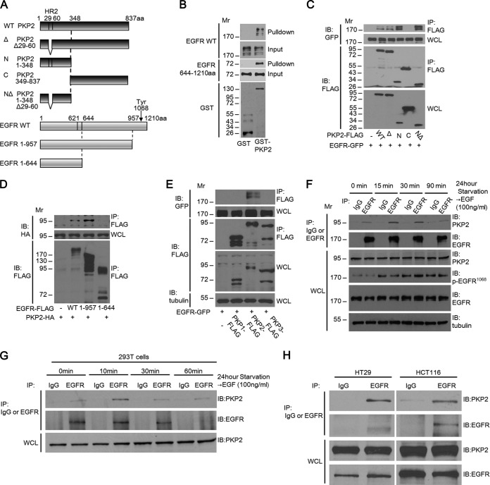
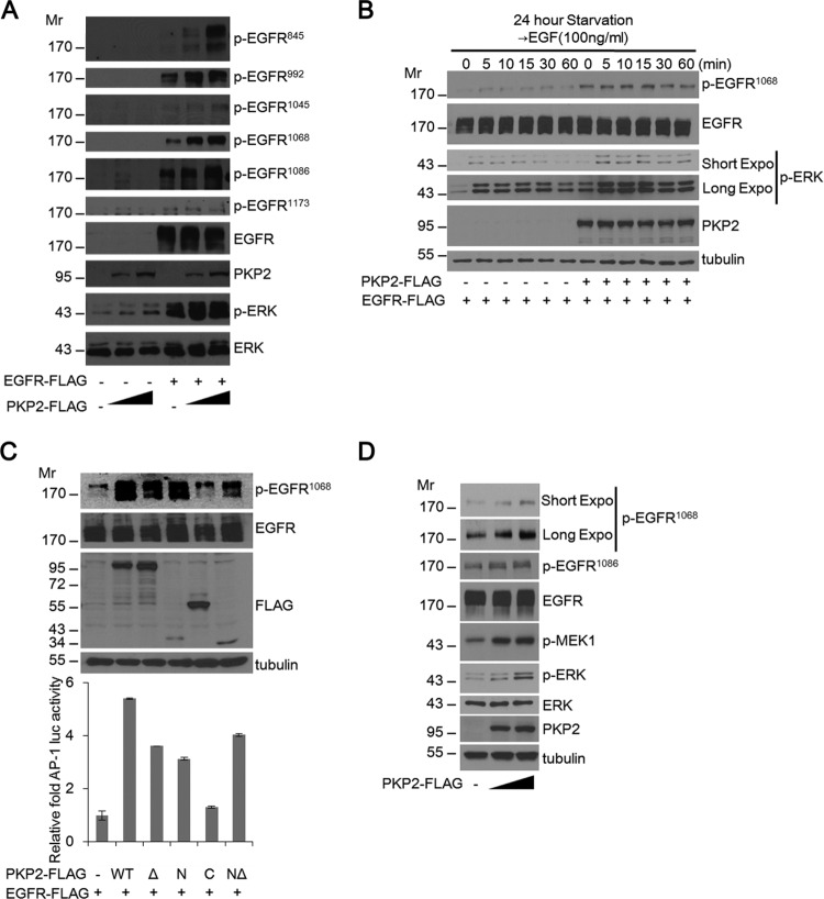
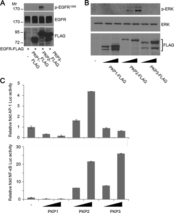
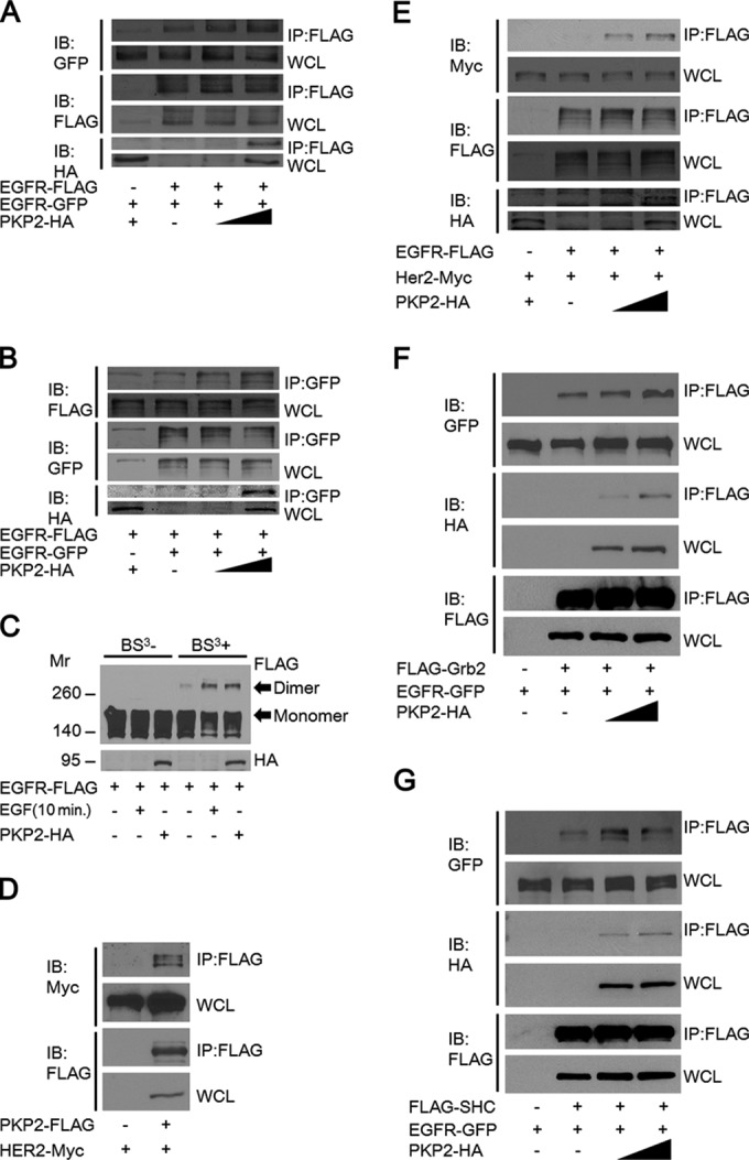
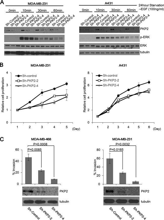
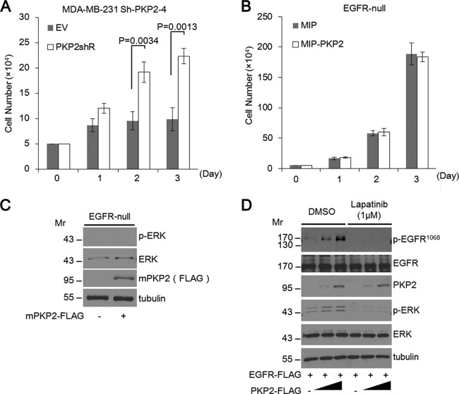
Epidermal growth factor (EGF) receptor (EGFR) has been implicated in tumor development and invasion. Dimerization and autophosphorylation of EGFR are the critical events for EGFR activation. However, the regulation of EGF-dependent and EGF-independent dimerization and phosphorylation of EGFR has not been fully understood. Here, we report that cytoplasmic protein plakophilin-2 (PKP2) is a novel positive regulator of EGFR signaling. PKP2 specifically interacts with EGFR via its N-terminal head domain. Increased PKP2 expression enhances EGF-dependent and EGF-independent EGFR dimerization and phosphorylation. Moreover, PKP2 knockdown reduces EGFR phosphorylation and attenuates EGFR-mediated signal activation, resulting in a significant decrease in proliferation and migration of cancer cells and tumor development. Our results indicate that PKP2 is a novel activator of the EGFR signaling pathway and a potential new drug target for inhibiting tumor growth.


Figures

FIG 1
FIG 1
PKP2 interacts with EGFR. (A) Schematic drawings of PKP2, EGFR, and the derivatives used in this work. A capital delta indicates an HR2 motif (aa 29 to 60) deletion. The location of EGFR Tyr1068 is indicated. Since the first 24-aa sequence of EGFR is a signal peptide sequence, the actual amino acid sequence of Tyr1068 is 1,092 aa. C, C terminus; N, N terminus; WT, wild type. (B) PKP2 directly interacted with the cytoplasmic region of EGFR. FLAG-tagged full-length EGFR or the cytoplasmic domain (aa 644 to 1210) of EGFR expressed in 293T cells was purified using immunoprecipitation and tested for binding to bacterially synthesized GST or GST-PKP2 on GST binding columns. Bound proteins were analyzed using Western blotting. (C) The N terminus of PKP2 is required for EGFR-PKP2 interaction. 293T cells were cotransfected with plasmids encoding EGFR-GFP and PKP2-FLAG or its mutants. Cell lysates were immunoprecipitated (IP) with anti-FLAG antibody and immunoblotted (IB) with GFP or FLAG. Whole-cell lysates (WCL) were also immunoblotted with the indicated antibodies. (D) The EGFR cytoplasmic domain interacted with PKP2 in 293T cells. 293T cells were cotransfected with plasmids encoding PKP2-HA and EGFR-FLAG or its mutants. Cell lysates were immunoprecipitated with anti-FLAG antibody and immunoblotted with HA or FLAG. Whole-cell lysates were also immunoblotted with HA. (E) PKP2 specifically interacts with EGFR among the members of the PKP family. 293T cells were cotransfected with plasmids encoding EGFR-GFP and PKP1, PKP2, or PKP3-FLAG. Cell lysates were immunoprecipitated with anti-FLAG antibody and immunoblotted with GFP or FLAG. Whole-cell lysates were also immunoblotted using the indicated antibodies. (F) Interaction of endogenous PKP2 and endogenous EGFR in A431 cells. A431 cells that had been serum starved (0.5% FBS) for 24 h were treated with EGF (100 ng/ml) at the indicated times, followed by immunoprecipitation with IgG or EGFR and immunoblotting with PKP2 (top rows) or EGFR (bottom rows) antibodies. Whole-cell lysates were also immunoblotted with the indicated antibodies. (G) 293T cells that had been serum starved (0.5% FBS) for 24 h were treated with EGF (100 ng/ml) at the indicated times, followed by immunoprecipitation with IgG or EGFR and immunoblotting with PKP2 (top rows) or EGFR (bottom rows) antibodies. Whole-cell lysates were also immunoblotted with the indicated antibodies. (H) Interaction of endogenous PKP2 and endogenous EGFR in HT29 (left column) and HCT116 (right column) cells. HT29 or HCT116 cells that had been serum starved (0.5% FBS) for 24 h were immunoprecipitated with IgG or EGFR and immunoblotted with PKP2 (top rows) or EGFR (bottom rows) antibodies. Whole-cell lysates were also immunoblotted with the indicated antibodies. Molecular weights are in thousands.
FIG 2
FIG 2
PKP2 expression enhances EGFR phosphorylation. (A) Serum-starved (1% FBS for 24 h) 293T cells were cotransfected with plasmids encoding empty vector or EGFR-FLAG and plasmid (0, 1, and 2 μg) expressing PKP2-FLAG as indicated. At 24 h after transfection, the cell lysates were then subjected to Western blotting using the indicated antibodies. (B) Expression of PKP2 increased EGFR phosphorylation and was enhanced after EGF stimulation in 293T cells. Mock-transfected or PKP2-expressing 293T cells were starved (1% FBS for 24 h), and EGF (100 ng/ml) was added for the indicated times. The cell lysates were then immunoblotted with the indicated antibodies. Expo, exposure. (C) N-terminal PKP2 expression enhanced EGFR phosphorylation and AP-1 luciferase activity. Serum-starved (1% FBS for 24 h) 293T cells were cotransfected with plasmids encoding AP-1-luc, EGFR-FLAG, and PKP2-FLAG and its mutants as indicated. At 24 h after transfection, AP-1 luciferase activities were analyzed. The protein levels of tubulin, EGFR, p-EGFR1068, and PKP2 and its mutants in the lysates are also shown. (D) PKP2 enhanced endogenous EGFR phosphorylation and increased downstream signal. Serum-starved (0.5% FBS for 24 h) A431 cells were transfected with plasmids (0 to 5 μg) encoding PKP2-FLAG. At 24 h after transfection, the cell lysates were examined using the indicated antibodies. Molecular weights are in thousands.
FIG 3
FIG 3
PKP2 is the only member of the plakophilin family capable of specifically activating EGFR signaling. (A) PKP2 specifically enhanced EGFR phosphorylation. Serum-starved (1% FBS for 24 h) 293T cells were cotransfected with plasmids encoding EGFR-FLAG and PKP1, PKP2, or PKP3-FLAG. At 24 h after transfection, the cell lysates were immunoblotted with the indicated antibodies. Molecular weights are in thousands. (B) At 24 h, serum-starved (1% FBS-containing DMEM) 293T cells were transfected with plasmids encoding increasing amounts of PKP1, PKP2, or PKP3-FLAG as indicated. At 24 h after transfection, the cell lysates were immunoblotted with the indicated antibodies. (C) 293T cells were transfected with plasmids encoding AP-1-Luc (upper panel) or NF-κB-Luc (lower panel) (50 ng), together with plasmids expressing PKP1, PKP2, or PKP3 (100 and 200 ng), as indicated. At 24 h after transfection, the luciferase activities were examined. Error bars represent the standard deviations (SD) of the means of the results of triplicate experiments.
FIG 4
FIG 4
EGFR dimerization is increased by PKP2 overexpression. (A and B) 293T cells were cotransfected with EGFR-GFP, EGFR-FLAG, and increasing amounts of PKP2-HA as indicated. Cell lysates were immunoprecipitated with anti-FLAG antibody (A) or anti-GFP antibody (B) and immunoblotted with antibodies as indicated. Whole-cell lysates were also immunoblotted with the indicated antibodies. (C) The level of EGFR dimerization was increased by PKP2 expression. Serum-starved (1% FBS for 24 h) 293T cells were cotransfected with EGFR-FLAG and PKP2-HA as indicated. As a control, EGF (10 ng/ml) was added for 10 min. Cell lysates with or without BS3 cross-linking were analyzed by Western blotting. Molecular weights are in thousands. (D) PKP2 interacted with HER2. 293T cells were cotransfected with HER2-Myc and PKP2-FLAG. Cell lysates were immunoprecipitated with anti-FLAG antibody and immunoblotted with FLAG or Myc antibodies. Whole-cell lysates were also immunoblotted with FLAG or Myc antibodies. (E) The level of EGFR-HER2 interaction was increased by PKP2 overexpression. 293T cells were cotransfected with EGFR-FLAG, HER2-Myc, and increasing amounts of PKP2-HA as indicated. Cell lysates were immunoprecipitated with anti-FLAG antibody and immunoblotted with antibodies as indicated. Whole-cell lysates were also immunoblotted with the indicated antibodies. (F) The level of EGFR-Grb2 interaction was increased by PKP2 overexpression. 293T cells were cotransfected with EGFR-GFP, FLAG-Grb2, and increasing amounts of PKP2-HA as indicated. Cell lysates were immunoprecipitated with anti-FLAG antibody and immunoblotted with antibodies as indicated. Whole-cell lysates were also immunoblotted with the indicated antibodies. (G) The level of EGFR-SHC interaction was increased by PKP2 overexpression. 293T cells were cotransfected with EGFR-GFP, FLAG-SHC, and increasing amounts of PKP2-HA as indicated. Cell lysates were immunoprecipitated with anti-FLAG antibody and immunoblotted with antibodies as indicated. Whole-cell lysates were also immunoblotted with antibodies as indicated.
FIG 5
FIG 5
PKP2 knockdown impairs EGF-induced responses. (A) Efficiency of PKP2 knockdown using siRNA and shRNA in 293T cells. The mRNA levels of PKP2 were visualized using RT-PCR. To assess the protein levels of PKP2 to test the efficiency of siRNAs and shRNAs, plasmids encoding PKP1 and PKP2 with siRNAs or shRNAs were transfected to 293T cells. At 48 h after transfection, the cell lysates were subjected to Western blot analysis. si-Control, control siRNA; sh-Control, control shRNA. (B) A431 cells were treated with control siRNA or si-PKP2-2 (twice, at 0 and 48 h). At 72 h after the first siRNA transfection, the media were changed to 0.5% FBS-containing DMEM. EGF was added at 98 h after the first siRNA transfection and was harvested at the indicated times. (C) A431 cells were transfected with plasmids encoding control shRNA, shPKP2-2, or shPKP2-4, followed by selection by puromycin. Selected cells were serum starved with 0.5% FBS-containing DMEM for 24 h, and EGF was added and harvested at the indicated times. (D) The ratios of tyrosine 1068-phosphorylated EGFR to total EGFR presented in panel C were quantified. (E) HCT116 cells were transfected with plasmids encoding control shRNA or shPKP2-4, followed by selection with puromycin. Selected cells were serum starved in DMEM with 1% FBS for 24 h, and then EGF was added and harvested at the indicated times. (F) MDA-MB-468 cells were transfected with plasmids encoding control shRNA or shPKP2-4, followed by selection with puromycin. Selected cells were serum starved with 0.5% FBS DMEM for 24 h, and EGF was added and harvested at the indicated times. Cell lysates were immunoprecipitated with anti-EGFR antibody and immunoblotted with antibodies as indicated. (G) A431 cells were transfected with plasmids encoding control shRNA or sh-PKP2-4, followed by selection with puromycin. Selected cells were serum starved with 0.5% FBS DMEM for 24 h, and EGF was added and harvested at the indicated times. Cell lysates were immunoprecipitated with anti-EGFR antibody and immunoblotted with antibodies as indicated. Whole-cell lysates were also immunoblotted as indicated. (H) Expression of PKP2 increased the interactions between EGFR and adopter molecules. Sh-PKP2-4-expressing A431 cells were transfected with plasmids encoding empty PKP2 or increasing amounts of Sh-PKP2-4-resistant PKP2 as indicated. Cell lysates were immunoprecipitated with IgG or anti-EGFR antibody and immunoblotted with antibodies as indicated. Whole-cell lysates were also immunoblotted as indicated. (I) The sh-PKP2-4-expressing A431 cells were transfected with plasmids encoding empty vector (EV) or sh-PKP2-4-resistant PKP2 (PKP2shR). At 24 h after starvation (0.5% FBS-containing DMEM), EGF was added and harvested at the indicated times. (J) Suppression of PKP2 expression reduces the EGFR dimerization rate. The short hairpin control or sh-PKP2-4-expressing A431 cells were transiently transfected with EGFR-GFP and empty vector or EGFR-FLAG as indicated. At 24 h after transfection, cell lysates were immunoprecipitated with FLAG and immunoblotted with antibodies as indicated. Whole-cell lysates were also immunoblotted as indicated. Molecular weights are in thousands.
FIG 6
FIG 6
Suppression of PKP2 expression reduces cell proliferation and invasion. (A) The short hairpin control (sh-Control)-, sh-PKP2-2-, or sh-PKP2-4-expressing MDA-MB-231 cells or A431 cells (left panel or right panel, respectively) were serum starved with 0.5% FBS-containing DMEM for 24 h. At the indicated times after EGF (100 ng/ml) treatment, cells were lysed and analyzed using the indicated antibodies. (B) The cell growth of short hairpin control (sh-Control)-, sh-PKP2-2-, or sh-PKP2-4-expressing MDA-MB-231 or MDA-MB-468 was monitored by an MTS assay (left panel or right panel, respectively). (C) Knockdown of PKP2 substantially impaired cell invasion. MDA-MB-468 cells (left) or MDA-MB-231 cells (right) were transfected with the short hairpin control (sh-Control), sh-PKP2-2, or sh-PKP2-4 and selected using puromycin. The selected cells were analyzed using the Matrigel invasion assay. The protein levels of PKP2 are also shown (bottom).
FIG 7
FIG 7
PKP2 functions directly through EGFR, and its effects are abolished in EGFR null cells. (A) The sh-PKP2-4-expressing MDA-MB-231 cells were transfected with plasmids encoding empty vector (EV) or sh-PKP2-4-resistant PKP2 (PKP2shR). After selection, these cells were monitored by trypan blue counting. (B) 32D cells stably expressing MIP or MIP-mouse PKP2 were monitored by trypan blue counting. (C) Mouse PKP2 was transiently expressed in 32D cells as indicated. Twenty-four hours after transfection, cell lysates were immunoblotted with the indicated antibodies. (D) Lapatinib treatment completely impaired the enhancement of PKP2-mediated EGFR phosphorylation. Serum-starved (1% FBS for 24 h) 293T cells were cotransfected with plasmids encoding EGFR-FLAG and PKP2-FLAG as indicated. The cells were treated with DMSO or lapatinib (final 1 μM) for 24 h. At 24 h after transfection, the cell lysates were subjected to Western blotting using the indicated antibodies. Molecular weights are in thousands.
FIG 8
FIG 8
PKP2 regulates breast cancer development in a mouse model. (A) Pieces of tumors were homogenized and subjected to Western blotting and analyzed using PKP2 and tubulin antibodies. Molecular weights are in thousands. (B) All tumors analyzed in this assay are shown. (n = 5; short hairpin control [sh-Control], sh-PKP2-2, and sh-PKP2-4). (C) Primary tumor growth was measured upon orthotopic injection of MDA-MB-231 cells with expression of the short hairpin control (sh-Control), sh-PKP2-2, or sh-PKP2-4. The experiment was terminated 10 weeks after injection (n = 5 per group per time point). Each time point shows the means ± standard errors of the means (SEM) of the results. (D) At 10 weeks after orthotopic implantation of MDA-MB-231 cells with or without PKP2 knockdown, lungs were stained by H&E. Representative H&E stains of lungs are shown. Arrows indicate metastatic foci. (E) Quantitative results of the lung metastasis analysis described for panel D.

Comment in

References

    1. Blume-Jensen P, Hunter T. 2001. Oncogenic kinase signalling. Nature 411:355–365. 10.1038/35077225 - DOI - PubMed
    1. Bogdan S, Klambt C. 2001. Epidermal growth factor receptor signaling. Curr. Biol. 11:R292–R295. 10.1016/S0960-9822(01)00167-1 - DOI - PubMed
    1. Hynes NE, Lane HA. 2005. ERBB receptors and cancer: the complexity of targeted inhibitors. Nat. Rev. Cancer 5:341–354. 10.1038/nrc1609 - DOI - PubMed
    1. Hynes NE, MacDonald G. 2009. ErbB receptors and signaling pathways in cancer. Curr. Opin. Cell Biol. 21:177–184. 10.1016/j.ceb.2008.12.010 - DOI - PubMed
    1. Yarden Y, Pines G. 2012. The ERBB network: at last, cancer therapy meets systems biology. Nat. Rev. Cancer 12:553–563. 10.1038/nrc3309 - DOI - PubMed

Publication types

MeSH terms

LinkOut - more resources