. 2015 Jan 1;24(1):1-8.
doi: 10.1093/hmg/ddu414. Epub 2014 Aug 11.

A murine model of neurofibromatosis type 2 that accurately phenocopies human schwannoma formation

Affiliations

A murine model of neurofibromatosis type 2 that accurately phenocopies human schwannoma formation

Jeffrey R Gehlhausen et al. Hum Mol Genet. .

Abstract

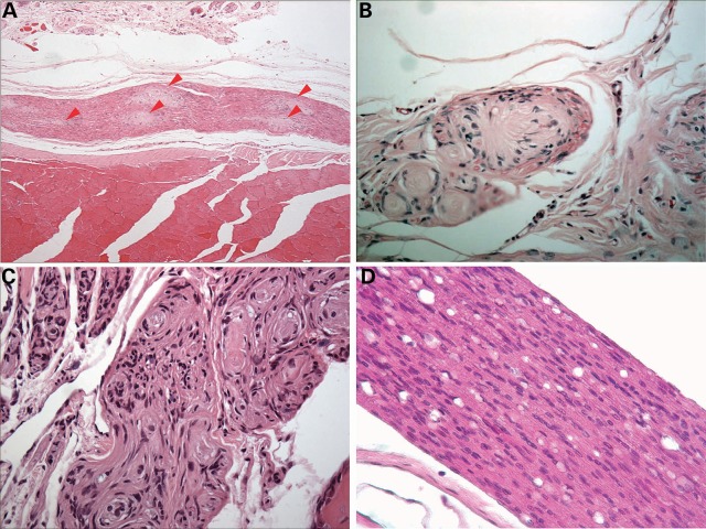
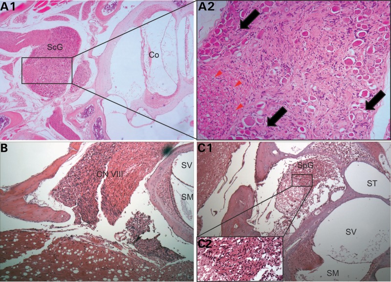
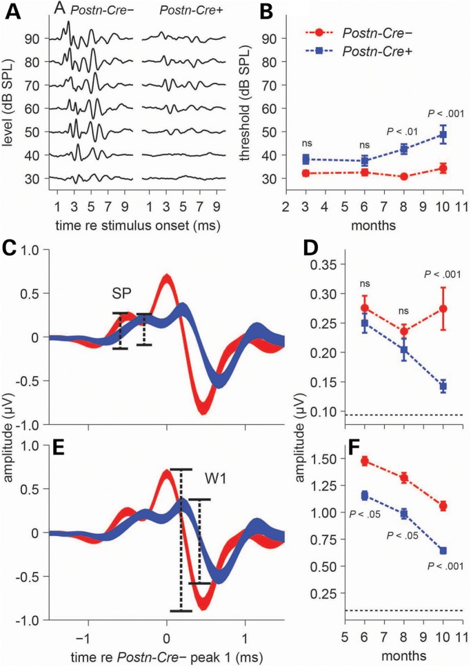
Neurofibromatosis type 2 (NF2) is an autosomal dominant genetic disorder resulting from germline mutations in the NF2 gene. Bilateral vestibular schwannomas, tumors on cranial nerve VIII, are pathognomonic for NF2 disease. Furthermore, schwannomas also commonly develop in other cranial nerves, dorsal root ganglia and peripheral nerves. These tumors are a major cause of morbidity and mortality, and medical therapies to treat them are limited. Animal models that accurately recapitulate the full anatomical spectrum of human NF2-related schwannomas, including the characteristic functional deficits in hearing and balance associated with cranial nerve VIII tumors, would allow systematic evaluation of experimental therapeutics prior to clinical use. Here, we present a genetically engineered NF2 mouse model generated through excision of the Nf2 gene driven by Cre expression under control of a tissue-restricted 3.9kbPeriostin promoter element. By 10 months of age, 100% of Postn-Cre; Nf2(flox/flox) mice develop spinal, peripheral and cranial nerve tumors histologically identical to human schwannomas. In addition, the development of cranial nerve VIII tumors correlates with functional impairments in hearing and balance, as measured by auditory brainstem response and vestibular testing. Overall, the Postn-Cre; Nf2(flox/flox) tumor model provides a novel tool for future mechanistic and therapeutic studies of NF2-associated schwannomas.


Figures

Figure 1.
Figure 1.
Postn-Cre; Nf2flox/flox mice develop schwannomas of the DRG and spinal nerves. (A and B) Dissected spinal cord of 15-month-old Cre-negative control (A) and Postn-Cre; Nf2flox/flox (B) mice, with Postn-Cre; Nf2flox/flox mice displaying diffusely enlarged DRG and spinal nerves (red arrowheads). (C and D) Hematoxylin and eosin (H&E) stain of the DRG from 7-month Cre-negative control and Postn-Cre; Nf2flox/flox mice, respectively. The whorls of Schwann cell proliferation observed in D are characteristic of schwannoma histology. Original magnification ×200. (E) Immunohistochemical S100 stain indicating tumors are comprised of mature Schwann cells. Original magnification ×400. (F) DRG volume quantitation of 15-month-old Cre-negative control mice (4) and Postn-Cre; Nf2flox/flox mice (6). P < 0.0001, unpaired Student's t-test with Welch's correction. (G) Kaplan–Meier survival analysis of Cre-negative control mice (7) and Postn-Cre;Nf2flox/flox mice (8). P < 0.001, log-rank (Mantel–Cox) test.
Figure 2.
Figure 2.
Schwannomas observed in Postn-Cre; Nf2flox/flox mice histologically resemble those observed in human patients. (A) H&E stain showing a multifocal pattern of schwannoma growth along a nerve (red arrowheads). Original magnification ×100. (B) Example of a plexiform schwannoma involving a nerve twig with peripheral axons. Original magnification ×400. (C) Pseudo-onion bulb formation of proliferating Schwann cells forming whorls around centrally located axons within a nerve. Original magnification ×200. (D) Diffuse spinal nerve hyperplasia observed in the nerve of a Postn-Cre; Nf2flox/flox mice. Original magnification ×400.
Figure 3.
Figure 3.
Postn-Cre; Nf2flox/flox mice develop schwannomas on CN VIII. (A1) H&E stain of a vestibular schwannoma located in Scarpa's ganglion (ScG). Original magnification ×100. In the 200× magnified inset image (A2), notice the Schwann cell proliferation displacing nerve cell bodies (black arrows) and pushing axons to the periphery of the nerve (red arrowheads). (B) Diffuse hypercellularity observed in CN VIII. Original magnification ×100. (C1) Schwann cell proliferation in the spiral ganglion (SpG), expanding and disrupting the architecture of the ganglion. Original magnification ×100. In the magnified (400×) inset C2, notice the dysplastic, irregularly spaced Schwann cells. Cochlea (Co), scala media (SM), scala vestibuli (SV) and scala tympani (ST).
Figure 4.
Figure 4.
Postn-Cre; Nf2flox/flox mice show deficits in both threshold and supra-threshold ABRs. (A) Representative ABR waveforms from individual Cre-negative (left) and Postn-Cre; Nf2flox/flox mice (right) at 10 months of age. (B) Time-course click-ABR study quantitating differences between Cre-negative and Postn-Cre; Nf2flox/flox mice. Significant threshold elevations were seen in Postn-Cre; Nf2flox/flox mice at 8 (P < 0.01) and 10 (P < 0.001) months of age (n = 15, 12, 15 and 19 mice for 3, 6, 8 and 10 months, respectively). Thresholds were averaged across animals (mean ± SEM). (C and D) The SP (C), dominated by inner-hair-cell receptor potentials and evident in the ABR waveform immediately preceding peak 1, is comparable in amplitude across groups at 6 and 8 months of age but is reduced in Postn-Cre; Nf2flox/flox mice at 10 months (P < 0.001) (D). (E and F) ABR wave 1 (W1) (E), representing the summed response of the cochlear nerve, is reduced in Postn-Cre; Nf2flox/flox mice compared with Cre-negative controls at all ages (P < 0.05, P < 0.05, P < 0.001) (F), indicating deficits in pre- and/or post-synaptic cochlear-nerve function. All statistical tests: two-way ANOVA with Bonferonni post hoc analysis. Thickness of waveforms (C and E) represents mean ± SEM. Amplitudes (D and F) represent high-level responses (across 60–90 dB SPL) averaged across animals (mean ± SEM). Horizontal dotted lines (D and F) at the base of the y-axis indicate the noise floor measurement.

References

    1. Asthagiri A.R., Parry D.M., Butman J.A., Kim H.J., Tsilou E.T., Zhuang Z., Lonser R.R. Neurofibromatosis type 2. Lancet. 2009;373:1974–1986. - PMC - PubMed
    1. Di Maio S., Akagami R. Prospective comparison of quality of life before and after observation, radiation, or surgery for vestibular schwannomas. J. Neurosurg. 2009;111:855–862. - PubMed
    1. Sharpless N.E., Depinho R.A. The mighty mouse: genetically engineered mouse models in cancer drug development. Nat. Rev. Drug. Discov. 2006;5:741–754. - PubMed
    1. Giovannini M., Robanus-Maandag E., Niwa-Kawakita M., van der Valk M., Woodruff J.M., Goutebroze L., Merel P., Berns A., Thomas G. Schwann cell hyperplasia and tumors in transgenic mice expressing a naturally occurring mutant NF2 protein. Genes Dev. 1999;13:978–986. - PMC - PubMed
    1. Giovannini M., Robanus-Maandag E., van der Valk M., Niwa-Kawakita M., Abramowski V., Goutebroze L., Woodruff J.M., Berns A., Thomas G. Conditional biallelic Nf2 mutation in the mouse promotes manifestations of human neurofibromatosis type 2. Genes Dev. 2000;14:1617–1630. - PMC - PubMed

Publication types