

Role of imaging in the staging and response assessment of lymphoma: consensus of the International Conference on Malignant Lymphomas Imaging Working Group
- PMID: 25113771
- PMCID: PMC5015423
- DOI: 10.1200/JCO.2013.53.5229
Role of imaging in the staging and response assessment of lymphoma: consensus of the International Conference on Malignant Lymphomas Imaging Working Group
Erratum in
-
ERRATUM.J Clin Oncol. 2016 Jul 20;34(21):2562. doi: 10.1200/JCO.2016.68.9265. J Clin Oncol. 2016. PMID: 27411915 Free PMC article. No abstract available.
Abstract
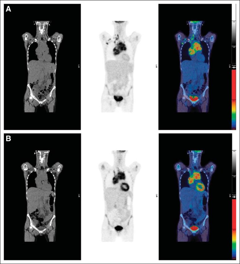
Purpose: Recent advances in imaging, use of prognostic indices, and molecular profiling techniques have the potential to improve disease characterization and outcomes in lymphoma. International trials are under way to test image-based response–adapted treatment guided by early interim positron emission tomography (PET)–computed tomography (CT). Progress in imaging is influencing trial design and affecting clinical practice. In particular, a five-point scale to grade response using PET-CT, which can be adapted to suit requirements for early- and late-response assessment with good interobserver agreement, is becoming widely used both in practice- and response-adapted trials. A workshop held at the 11th International Conference on Malignant Lymphomas (ICML) in 2011 concluded that revision to current staging and response criteria was timely.
Methods: An imaging working group composed of representatives from major international cooperative groups was asked to review the literature, share knowledge about research in progress, and identify key areas for research pertaining to imaging and lymphoma.
Results: A working paper was circulated for comment and presented at the Fourth International Workshop on PET in Lymphoma in Menton, France, and the 12th ICML in Lugano, Switzerland, to update the International Harmonisation Project guidance regarding PET. Recommendations were made to optimize the use of PET-CT in staging and response assessment of lymphoma, including qualitative and quantitative methods.
Conclusion: This article comprises the consensus reached to update guidance on the use of PET-CT for staging and response assessment for [18F]fluorodeoxyglucose-avid lymphomas in clinical practice and late-phase trials.
Conflict of interest statement
Authors' disclosures of potential conflicts of interest and author contributions are found at the end of this article.
Figures
Comment in
-
Quantitative positron emission tomography imaging for response assessment of [18F]fluorodeoxyglucose-avid lymphomas: should we report standard uptake value confidence limits?J Clin Oncol. 2015 Apr 1;33(10):1218-9. doi: 10.1200/JCO.2014.59.6833. Epub 2015 Feb 17. J Clin Oncol. 2015. PMID: 25691673 No abstract available.
-
Do not abandon the bone marrow biopsy yet in diffuse large B-cell lymphoma.J Clin Oncol. 2015 Apr 1;33(10):1217. doi: 10.1200/JCO.2014.58.7360. Epub 2015 Feb 17. J Clin Oncol. 2015. PMID: 25691675 No abstract available.
-
Value of positron emission tomography in diagnosing subcutaneous panniculitis-like T-cell lymphoma.J Clin Oncol. 2015 Apr 1;33(10):1216-7. doi: 10.1200/JCO.2014.59.8193. Epub 2015 Feb 17. J Clin Oncol. 2015. PMID: 25691676 No abstract available.
-
Reply to B. Bennani-Baiti et al, H.J.A. Adams et al, E. Laffon et al, and E.A. Hawkes et al.J Clin Oncol. 2015 Apr 1;33(10):1221-3. doi: 10.1200/JCO.2014.59.9373. Epub 2015 Feb 17. J Clin Oncol. 2015. PMID: 25691678 No abstract available.
-
Fallacy of Quantifying Lymphoma Activity by Scaling to the Liver in [18F]Fluorodeoxyglucose Positron Emission Tomography (Deauville criteria).J Clin Oncol. 2015 Dec 1;33(34):4120-1. doi: 10.1200/JCO.2015.63.1382. Epub 2015 Aug 24. J Clin Oncol. 2015. PMID: 26304889 No abstract available.
-
Reply to G. Keramida et al.J Clin Oncol. 2015 Dec 1;33(34):4121-2. doi: 10.1200/JCO.2015.63.2125. Epub 2015 Aug 24. J Clin Oncol. 2015. PMID: 26304890 No abstract available.
References
-
- A predictive model for aggressive non-Hodgkin's lymphoma: The International Non-Hodgkin's Lymphoma Prognostic Factors Project. N Engl J Med. 1993;329:987–994. - PubMed
-
- Solal-Céligny P, Roy P, Colombat P, et al. Follicular lymphoma international prognostic index. Blood. 2004;104:1258–1265. - PubMed
-
- Hoster E, Dreyling M, Klapper W, et al. A new prognostic index (MIPI) for patients with advanced-stage mantle cell lymphoma. Blood. 2008;111:558–565. - PubMed
-
- Younes A. Early-stage Hodgkin's lymphoma: In pursuit of perfection. J Clin Oncol. 2012;30:895–896. - PubMed
-
- Rosenwald A, Wright G, Chan WC, et al. The use of molecular profiling to predict survival after chemotherapy for diffuse large-B-cell lymphoma. N Engl J Med. 2002;346:1937–1947. - PubMed
Publication types
MeSH terms
Substances
LinkOut - more resources
Full Text Sources
Other Literature Sources
Medical
