. 2014 Aug 25;211(9):1905-18.
doi: 10.1084/jem.20132577. Epub 2014 Aug 11.

Interplay between regulatory T cells and PD-1 in modulating T cell exhaustion and viral control during chronic LCMV infection

Affiliations

Interplay between regulatory T cells and PD-1 in modulating T cell exhaustion and viral control during chronic LCMV infection

Pablo Penaloza-MacMaster et al. J Exp Med. .

Abstract

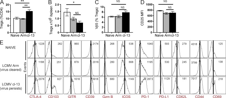
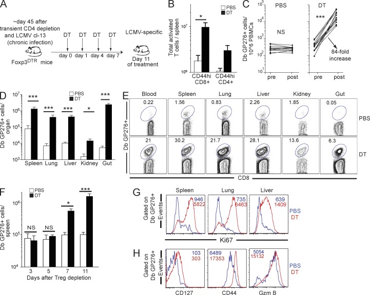
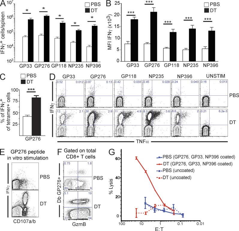
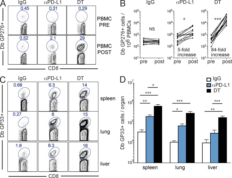
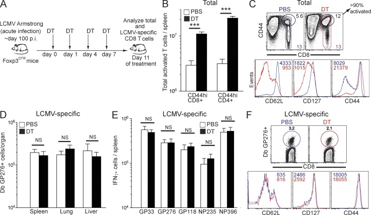
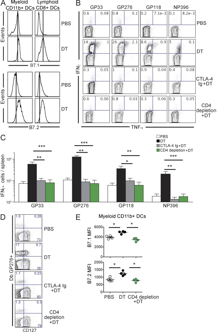
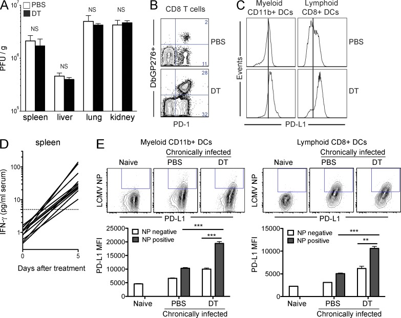
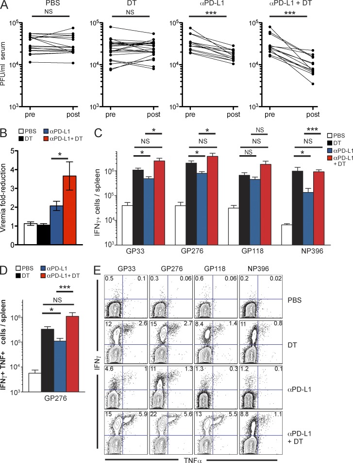
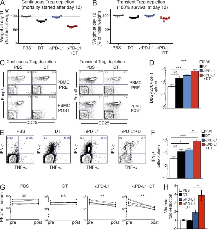
Regulatory T (T reg) cells are critical for preventing autoimmunity mediated by self-reactive T cells, but their role in modulating immune responses during chronic viral infection is not well defined. To address this question and to investigate a role for T reg cells in exhaustion of virus-specific CD8 T cells, we depleted T reg cells in mice chronically infected with lymphocytic choriomeningitis virus (LCMV). T reg cell ablation resulted in 10-100-fold expansion of functional LCMV-specific CD8 T cells. Rescue of exhausted CD8 T cells was dependent on cognate antigen, B7 costimulation, and conventional CD4 T cells. Despite the striking recovery of LCMV-specific CD8 T cell responses, T reg cell depletion failed to diminish viral load. Interestingly, T reg cell ablation triggered up-regulation of the molecule programmed cell death ligand-1 (PD-L1), which upon binding PD-1 on T cells delivers inhibitory signals. Increased PD-L1 expression was observed especially on LCMV-infected cells, and combining T reg cell depletion with PD-L1 blockade resulted in a significant reduction in viral titers, which was more pronounced than that upon PD-L1 blockade alone. These results suggest that T reg cells effectively maintain CD8 T cell exhaustion, but blockade of the PD-1 inhibitory pathway is critical for elimination of infected cells.


Figures

Figure 1.
Figure 1.
T reg cells have an activated phenotype during chronic viral infection. Analysis was done on splenocytes from naive uninfected mice, mice infected with LCMV Arm, and analyzed at least 100 d after acute infection and LCMV cl-13 chronically infected mice (mice transiently depleted of CD4 T cells and infected with LCMV cl-13, with analysis done at least 40 d after infection). (A) Percentage of T reg cells (Foxp3+) among CD4 T cells. (B) Absolute number of T reg cells in spleen. (C) Frequency of Vβ5+ cells among T reg cells. Mice infected with LCMV cl-13 and analyzed 21 d later were included as positive controls (22–40% Vβ5+ cells). (D) Graph shows MFI of CD25 expression on T reg cells. (E) Histograms show representative expression of different markers by T reg cells and numbers represent MFI. Data are a compilation of 4 experiments (A and B) or show representative results from 3 experiments (C–E; n = 3–4 mice per group). Error bars indicate SEM. Non-parametric Mann Whitney test, where *, P < 0.05; **, P < 0.01; NS = not significant.
Figure 2.
Figure 2.
Exhausted CD8 T cells expand and undergo phenotypic changes upon T reg cell ablation. (A) Experimental outline. (B) Total numbers of activated T cells in the spleen of PBS-treated or T reg cell–depleted (DT) mice chronically infected with LCMV. (C) Number of LCMV-specific (Db GP276) CD8 T cells among PBMCs before and after DT treatment. (D and E) Absolute numbers (D) and frequency (E) of LCMV-specific (Db GP276) CD8 T cells within total CD8 T cell population. (F) Absolute numbers of LCMV DbGP276-specific CD8 T cells in the spleen at different days after T reg cell ablation. (G) Proliferative activity of LCMV-specific (Db-GP276) CD8 T cells. Numbers show MFI of Ki67 expression. (H) Phenotype of LCMV-specific (Db GP276) CD8 T cells in spleen. Numbers show MFI of different CD8 T cell markers. Data are a compilation of 5 independent experiments with 3–5 mice per group. Error bars indicate SEM. Non-parametric Mann Whitney test, where *, P < 0.05; ***, P < 0.001; NS = not significant.
Figure 3.
Figure 3.
LCMV-specific CD8 T cells regain effector function upon T reg cell ablation in chronically infected mice. LCMV chronically infected Foxp3DTR knock-in mice were depleted of T reg cells for 10 d by DT administration as in Fig. 2. (A) Absolute numbers of CD8 T cells in spleen producing IFN-γ. (B) MFI of IFN-γ production by CD8 T cells after in vitro restimulation with various LCMV peptides. (C) Percentage among DbGP276-specific cells that express IFN-γ in spleen. (D) Frequency of CD8 T cells producing both IFN-γ and TNF after in vitro restimulation with LCMV peptides. (E) Degranulation and surface expression of CD107 a/b in CD8 T cells after in vitro restimulation with GP276 LCMV peptide. (F) Granzyme B expression on LCMV DbG276-specific CD8 T cells in spleen. (G) Ex vivo cytotoxic activity of splenic CD8 T cells measured by 51Cr release from MC57 target cells unpulsed (control) or pulsed with a mix of LCMV peptides (GP33, GP276, and NP396). Data are a compilation of 5 independent experiments with 3–5 mice per group. Error bars indicate SEM. Non-parametric Mann Whitney test, where *, P < 0.05; ***, P < 0.001.
Figure 4.
Figure 4.
Rescue of LCMV-specific CD8 T cell responses by T reg cell ablation is greater than by blockade of the PD-1 pathway. LCMV chronically infected Foxp3DTR knock-in mice were depleted of T reg cells for 10 d by DT administration as in Fig. 2 or mice received 3 doses of PD-L1 blocking antibody, every 3 d. (A and B) Dot plots (A) show frequency in the same mouse and graphs (B) show number of LCMV-specific (Db GP276) CD8 T cells among PBMCs before and after treatment. (C and D) Dot plots show frequency (C) and graphs show number (D) of LCMV-specific (Db GP33) CD8 T cells in different organs 11 d after treatment. Data are a compilation of 3 independent experiments with 3–5 mice per group. Error bars indicate SEM. Non-parametric Mann Whitney test, where *, P < 0.05; **, P < 0.01; ***, P < 0.001; NS = not significant.
Figure 5.
Figure 5.
Cognate antigen is necessary for activation of antiviral T cells after T reg cell depletion. (A) Experimental outline. (B) Total numbers of activated T cells in the spleen of PBS-treated or T reg cell–depleted (DT) mice 100 d after infection with LCMV Arm. (C) Phenotype of total CD8 T cells in spleen. (D) Absolute numbers of LCMV-specific (Db GP276) CD8 T cells. (E) Absolute numbers of cells in the spleen that produce IFN-γ after in vitro restimulation with various LCMV peptides. (F) Phenotype of splenic LCMV-specific (Db GP276) CD8 T cells. A representative of 3 independent experiments is shown, with 5–6 mice per group. Error bars indicate SEM. Non-parametric Mann Whitney test, where ***, P < 0.001; NS = not significant.
Figure 6.
Figure 6.
Co-stimulation and CD4 T cells are required for the rescue of exhausted CD8 T cells upon T reg cell depletion. Foxp3DTR knock-in mice chronically infected with LCMV cl-13 received DT for 10 d (as in Fig. 2). CTLA-4 Ig was administered every other day during T reg cell depletion starting at day −2, and CD4 T cells were depleted upon administration of CD4 antibody on days −2 and −1 of T reg cell ablation. (A) Representative dot plots show expression of B7.1 and B7.2 on splenic DCs after 8 d of T reg cell ablation. (B and C) Frequency of splenic CD8 T cells that co-express IFN-γ and TNF (B) and absolute numbers of CD8 T cells that produce IFN-γ (C) after in vitro restimulation with LCMV peptides. (D) Frequency of LCMV-specific (Db-GP276) and CD127-expressing splenic CD8 T cells. (E) Graphs show B7.1 and B7.2 MFI on CD11b+ splenic DCs after 8 d of T reg cell ablation preceded or not by conventional CD4 T cell depletion. The data are representative of 3 independent experiments with 3–5 mice per group. Error bars indicate SEM. Non-parametric Mann Whitney test, where *, P < 0.05; **, P < 0.01; ***, P < 0.001.
Figure 7.
Figure 7.
Inability to clear virus coincides with PD-L1 up-regulation. Foxp3DTR knock-in mice chronically infected with LCMV received DT for 10 d (as in Fig. 2). (A) Viral load after 10 d of T reg cell depletion. (B) Dot plots show PD-1 expression on DbGP276-specific CD8 T cells. (C) PD-L1 expression on splenic DCs after 7 d of T reg cell ablation. (D) Graph shows IFN-γ in serum before and at day 5 after initiation of DT treatment. Dashed line indicates the limit of detection of this assay. (E) Representative dot plots show intracellular staining of LCMV nucleoprotein (NP) and PD-L1 expression on splenic DCs. Staining of DCs in uninfected mice is shown as a control for specificity of NP staining and for basal PD-L1 expression. PD-L1 expression is shown as MFI on infected (NP positive) or noninfected (NP negative) DCs. Data are a compilation of 3 experiments (A) or show representative results of 2–3 experiments (B–E) with 3–6 mice per group. Error bars indicate SEM. (A) Non-parametric Mann Whitney test, where NS = not significant. (D) Wilcoxon matched pairs test, where *, P < 0.05. (E) Student’s t test, where **, P < 0.01; ***, P < 0.001.
Figure 8.
Figure 8.
Essential role for PD-L1 blockade in viral control after T reg cell depletion. Foxp3DTR knock-in mice chronically infected with LCMV received DT for 10 d (as in Fig. 2) in combination with PD-L1 blocking antibody. PD-L1 antibody was administered on days 1, 4, and 7 of DT treatment. (A) Viral titers in serum before and after treatment. (B) Fold reduction in serum viral titer after treatment. (C) Absolute numbers of splenic CD8 T cells that produce IFN-γ after in vitro restimulation with LCMV peptides. (D) Absolute numbers of CD8 T cells co-expressing IFN-γ and TNF after in vitro restimulation with GP276 LCMV peptide. (E) Representative dot plots showing frequency of CD8 T cells producing cytokines as in D. Data are a compilation (A and B) or show representative results of 3 independent experiments, with 4–6 mice per group. Error bars indicate SEM. Non-parametric Mann Whitney test, where *, P < 0.05; ***, P < 0.001; NS = not significant.
Figure 9.
Figure 9.
Transient T reg cell depletion improves CD8 T cell rescue and viral control when combined with PD-L1 blockade without causing overt disease in LCMV chronically infected mice. (A) Foxp3DTR knock-in mice chronically infected with LCMV received continuous DT (on days 0, 1, 4, and 7) and/or PD-L1 antibody (on days 1, 4, and 7), as in Fig. 8. Mice weight after 11 d of treatment, relative to initial weight before treatment. (B) As in A, but mice received transient DT treatment (on days 0, 1, and 4). Mice weight after 12 d of treatment. (C) Representative dot plots show frequency of T reg cells in PBMC before and after continuous or transient DT treatment. (D) Absolute numbers of LCMV-specific (Db GP276) CD8 T cells within the total splenic CD8 T cell population. (E and F) Frequency of CD8 T cells producing both IFN-γ and TNF (E) and absolute numbers of CD8 T cells in the spleen that produce IFN-γ after in vitro restimulation with LCMV peptide GP276 (F). (G) Viral titers in serum before and after treatment. (H) Fold reduction in serum viral titer after treatment. A representative of 3 independent experiments is shown, with 6 mice per group. Error bars indicate SEM. Non-parametric Mann Whitney test, where *, P < 0.05; **, P < 0.01; ***, P < 0.001; NS = not significant.

Similar articles

Cited by

References

    1. Ahmed, R., Salmi A., Butler L.D., Chiller J.M., and Oldstone M.B.. 1984. Selection of genetic variants of lymphocytic choriomeningitis virus in spleens of persistently infected mice. Role in suppression of cytotoxic T lymphocyte response and viral persistence. J. Exp. Med. 160:521–540 10.1084/jem.160.2.521 - DOI - PMC - PubMed
    1. Aubert, R.D., Kamphorst A.O., Sarkar S., Vezys V., Ha S.J., Barber D.L., Ye L., Sharpe A.H., Freeman G.J., and Ahmed R.. 2011. Antigen-specific CD4 T-cell help rescues exhausted CD8 T cells during chronic viral infection. Proc. Natl. Acad. Sci. USA. 108:21182–21187 10.1073/pnas.1118450109 - DOI - PMC - PubMed
    1. Barber, D.L., Wherry E.J., Masopust D., Zhu B., Allison J.P., Sharpe A.H., Freeman G.J., and Ahmed R.. 2006. Restoring function in exhausted CD8 T cells during chronic viral infection. Nature. 439:682–687 10.1038/nature04444 - DOI - PubMed
    1. Belkaid, Y., and Tarbell K.. 2009. Regulatory T cells in the control of host-microorganism interactions (*). Annu. Rev. Immunol. 27:551–589 10.1146/annurev.immunol.021908.132723 - DOI - PubMed
    1. Blackburn, S.D., Shin H., Haining W.N., Zou T., Workman C.J., Polley A., Betts M.R., Freeman G.J., Vignali D.A., and Wherry E.J.. 2009. Coregulation of CD8+ T cell exhaustion by multiple inhibitory receptors during chronic viral infection. Nat. Immunol. 10:29–37 10.1038/ni.1679 - DOI - PMC - PubMed

Publication types

MeSH terms

Substances