. 2014 Jul 30;5(14):5535-46.
doi: 10.18632/oncotarget.2137.

Tumor suppressor p53 negatively regulates glycolysis stimulated by hypoxia through its target RRAD

Affiliations

Tumor suppressor p53 negatively regulates glycolysis stimulated by hypoxia through its target RRAD

Cen Zhang et al. Oncotarget. .

Abstract

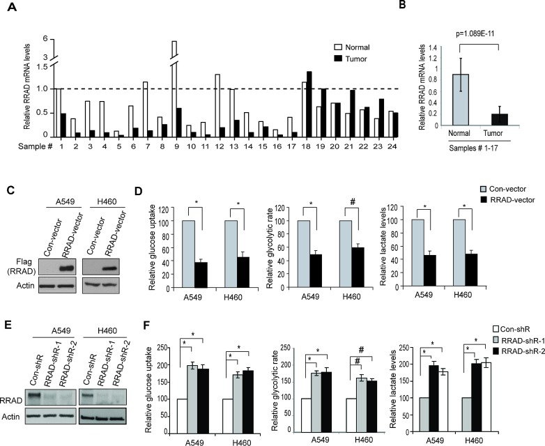
Cancer cells display enhanced glycolysis to meet their energetic and biosynthetic demands even under normal oxygen concentrations. Recent studies have revealed that tumor suppressor p53 represses glycolysis under normoxia as a novel mechanism for tumor suppression. As a common microenvironmental stress for tumors, hypoxia drives the metabolic switch from the oxidative phosphorylation to glycolysis, which is crucial for survival and proliferation of cancer cells under hypoxia. The p53's role and mechanism in regulating glycolysis under hypoxia is poorly understood. Here, we found that p53 represses hypoxia-stimulated glycolysis in cancer cells through RRAD, a newly-identified p53 target. RRAD expression is frequently decreased in lung cancer. Ectopic expression of RRAD greatly reduces glycolysis whereas knockdown of RRAD promotes glycolysis in lung cancer cells. Furthermore, RRAD represses glycolysis mainly through inhibition of GLUT1 translocation to the plasma membrane. Under hypoxic conditions, p53 induces RRAD, which in turn inhibits the translocation of GLUT1 and represses glycolysis in lung cancer cells. Blocking RRAD by siRNA greatly abolishes p53's function in repressing glycolysis under hypoxia. Taken together, our results revealed an important role and mechanism of p53 in antagonizing the stimulating effect of hypoxia on glycolysis, which contributes to p53's function in tumor suppression.


Conflict of interest statement

The authors declare no conflict of interest.

Figures

Figure 1
Figure 1. p53 reduces glucose uptake, the glycolytic rate and lactate production in human lung cancer cells under hypoxic conditions
(A) Hypoxia activated p53 in human lung A549 and H460 cells. The p53 wild-type A549-con-shR and H460-con-shR cells, as well as A549-p53-shR and H460-p53-shR cells with stable p53 knockdown by shRNA vectors were treated with hypoxia (0.1% O2) for 24 h before assays. Two different shRNA vectors against p53 were employed. The levels of p53 were measured by Western-blot assays. (B-D) Knockdown of p53 increased the glucose uptake (B), the glycolytic rate (C) and lactate production (D) in A549 and H460 cells under both normoxic and hypoxic conditions. Data are presented as mean value ± SD (n=3). #: p<0.05; * p<0.01 (Student's t tests).
Figure 2
Figure 2. RRAD negatively regulates glucose uptake, the glycolytic rate and lactate production in human lung cancer cells
(A) The relative RRAD mRNA expression in human lung cancer samples and their matched adjacent non-tumor tissues (n=24). The mRNA levels of RRAD were measured in 24 human lung cancer samples and their matched adjacent non-tumor tissues (Origene, Rockville) by Taqman real-time PCR assays. The mRNA levels of RRAD were normalized with Actin. The relative RRAD mRNA level in the adjacent non-tumor tissue of the cancer sample #1 was designated as 1. (B) The relative mean value of RRAD mRNA in human lung cancer samples #1-17 and their matched adjacent non-tumor lung tissues. Data are presented as mean value ± SD (n=17). P=1.089E-11. (C) Western-blot analysis of the ectopic RRAD expression in A549 and H460 cells stably transduced with pLPCX-RRAD-Flag retroviral vectors (RRAD) or control vectors (Con). (D) Ectopic RRAD expression reduced glucose uptake, the glycolytic rate and lactate production in A549 and H460 cells. (E) RRAD knockdown by shRNA in A549 and H460 cells detected by Western-blot assays. Cells were stably transduced with 2 different shRNA vectors against RRAD (RRAD-shR) or control shRNA (Con-shR). (F) Knockdown of endogenous RRAD by shRNA increased the glucose uptake, the glycolytic rate and lactate production in A549 and H460 cells. Data are presented as mean value ± SD (n=3). #: p<0.05; * p<0.01 (Student's t tests).
Figure 3
Figure 3. RRAD negatively regulates GLUT1 translocation to the plasma membrane (PM) in cells
(A) Ectopic RRAD expression reduced GLUT1 translocation to the PM in A549 and H460 cells as analyzed by Western-blot assays. The PM protein Na+/K+ ATPase was used as an internal control. The ER membrane protein Calnexin was used to exclude the contamination of PM by the other membrane fractions. (B) RRAD knockdown by shRNA promoted GLUT1 translocation to the PM in A549 and H460 cells. (C) Ectopic expression of RRAD or knockdown of endogenous RRAD did not change the mRNA levels of GLUT1 in A549 and H460 cells. The mRNA levels of GLUT1 were measured by Taqman real-time PCR, and normalized with Actin. (D) Ectopic RRAD expression reduced Myc-GLUT1 translocation to cell surface (left panel), whereas RRAD knockdown by shRNA promoted Myc-GLUT1 translocation to cell surface (right panel) in A549 and H460 cells analyzed in a flow cytometer. Relative Myc-GLUT1 levels on cell surface were calculated after normalization with the total Myc-GLUT1 levels in cells. Cells were transduced with pLPCX-Myc-GLUT1 vectors or control pLPCX vectors 48 h before assays. (E) Ectopic RRAD expression reduced Myc-GLUT1 translocation to the cell surface (left panels), whereas RRAD knockdown by shRNA promoted Myc-GLUT1 translocation to the cell surface (right panels) in A549 and H460 cells analyzed by IF staining with an anti-Myc antibody. (F) GLUT1 knockdown largely abolished the inhibitory effect of RRAD overexpression on glucose uptake, the glycolytic rate and lactate production in A549 and H460 cells. Cells with stable ectopic RRAD overexpression or control cells were transfected with 2 different GLUT1 siRNA (GLUT1-siR) or control siRNA for 24 h before assays. GLUT1 knockdown was confirmed by Western-blot assays (left panels). Data are presented as mean value ± SD (n=3). * p<0.01 (Student's t tests).
Figure 4
Figure 4. p53 induces RRAD expression under hypoxic conditions
(A, B) Activated p53 induced RRAD expression at both mRNA (A) and protein (B) levels under hypoxic conditions in A549 and H460 cells. A549-con-shR, A549-p53-shR, H460-con-shR and H460-p53-shR cells were treated with hypoxia for 18 or 24 h before mRNA and protein levels of RRAD were measured by real-time PCR (A) and Western-blot assays (B), respectively. The mRNA levels of RRAD were normalized with actin. p53 target MDM2 was used as a positive control for p53 activation. Two different p53 shRNA vectors were used and very similar results were obtained. (C) p53 bound to the p53 DNA binding element (p53 BE) in the RRAD promoter under hypoxic conditions detected by ChIP assays. Upper left panel: The consensus DNA sequences for the p53 BE. N, any nucleotide; Pu, purine; Py, pyramidine. Lower left panel: The p53 BE in human RRAD promoter. Number indicates the nucleotide position relative to the ATG site (+1). Right panel: ChIP analysis with the p53 antibody (DO-1) in A549-con-shR and A549-p53-shR cells treated with hypoxia for 18 h. The non-specific (NS) DNA fragment in the RRAD promoter (−1522 to −1244) which does not contain any potential p53 BE was used as a negative control. The p53 BE in MDM2 promoter served as a positive control.
Figure 5
Figure 5. RRAD mediates p53's function in negative regulation of glycolysis under hypoxic conditions
(A) Ectopic RRAD expression reduced glucose uptake, the glycolytic rate and lactate production stimulated by hypoxia in A549 and H460 cells. Cells with stable ectopic RRAD expression or control cells were treated with hypoxia for 24 h before assays. (B, C) RRAD mediated p53's function in repressing glucose uptake, the glycolytic rate and lactate production stimulated by hypoxia in A549 (B) and H460 (C) cells. Cells transduced with control or RRAD shRNA vectors together with or without p53 shRNA vectors were treated with hypoxia for 24 h before assays. Data were presented as mean value ± SD (n=3). #: p<0.05; * p<0.01 (Student's t tests).
Figure 6
Figure 6. p53 negatively regulates GLUT1 translocation to the plasma membrane (PM) through RRAD under hypoxic conditions
(A) Ectopic expression of RRAD reduced GLUT1 translocation to the PM under hypoxia in A549 cells as measured by Western-blot analysis. Cells with stable ectopic RRAD expression and control cells were treated with hypoxia for 24 h before assays. (B) Ectopic RRAD expression reduced Myc-GLUT1 translocation to the cell surface in A549 cells under hypoxia as measured by a flow cytometer. Cells were transfected with Myc-GLUT1 vectors and then treated with hypoxia for 24 h before assays. The levels of Myc-GLUT1 on the cell surface were detected by a flow cytometer, and normalized with the total levels of Myc-GLUT1 in cells. (C) p53 inhibited GLUT1 translocation to the PM under hypoxia in A549 cells as measured by Western-blot analysis. A549-con-shR and A549-p53-shR cells were treated with hypoxia for 24 h. (D) p53 inhibited Myc-GLUT1 translocation to the PM under hypoxia in A549 cells. (E, F) RRAD mediated p53's function in inhibition of GLUT1 translocation to the PM under hypoxia. A549-con-shR and A549-p53-shR cells were transduced with RRAD shRNA or control shRNA, followed by hypoxia treatment for 24 h before assays. (E) Western-blot analysis of the levels of endogenous GLUT1 on the plasma membrane. (F) The levels of Myc-GLUT1 on the cell surface measured by a flow cytometer. Data are presented as mean value ± SD (n=3). * p<0.01 (Student's t tests). (G) Schematic depicting that p53 negatively regulates glycolysis through the RRAD/GLUT1 signaling under hypoxia.

Similar articles

Cited by

References

    1. Warburg O. On the origin of cancer cells. Science. 1956;123(3191):309–314. - PubMed
    1. Gottlieb E, Vousden KH. p53 regulation of metabolic pathways. Cold Spring Harb Perspect Biol. 2010;2(4):a001040. - PMC - PubMed
    1. Cairns RA, Harris IS, Mak TW. Regulation of cancer cell metabolism. Nat Rev Cancer. 2011;11(2):85–95. - PubMed
    1. Christofk HR, Vander Heiden MG, Harris MH, Ramanathan A, Gerszten RE, Wei R, Fleming MD, Schreiber SL, Cantley LC. The M2 splice isoform of pyruvate kinase is important for cancer metabolism and tumour growth. Nature. 2008;452(7184):230–233. - PubMed
    1. Vousden KH, Prives C. Blinded by the Light: The Growing Complexity of p53. Cell. 2009;137(3):413–431. - PubMed

Publication types