

Autophagy protects C. elegans against necrosis during Pseudomonas aeruginosa infection
- PMID: 25114220
- PMCID: PMC4151725
- DOI: 10.1073/pnas.1405032111
Autophagy protects C. elegans against necrosis during Pseudomonas aeruginosa infection
Abstract
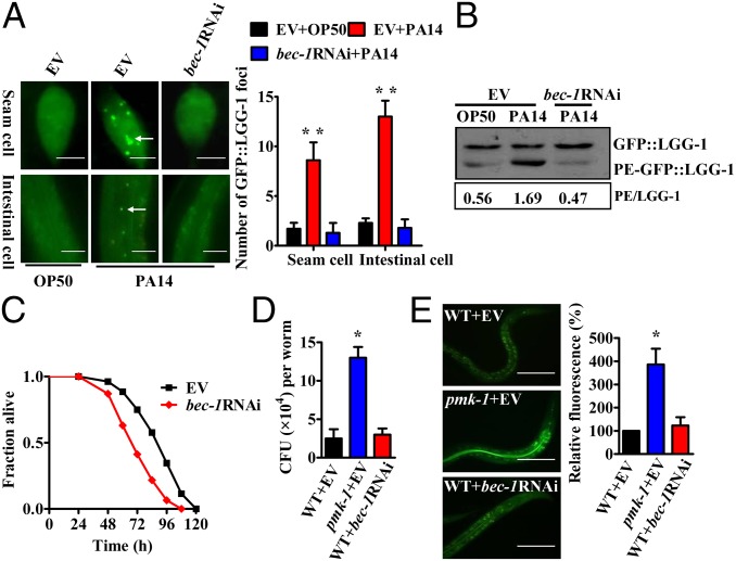
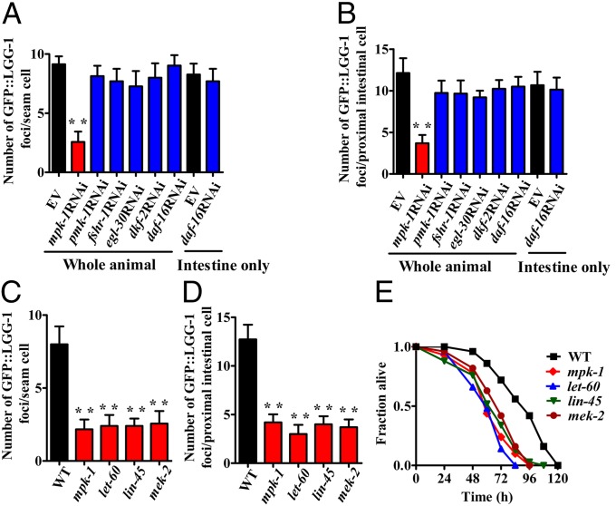
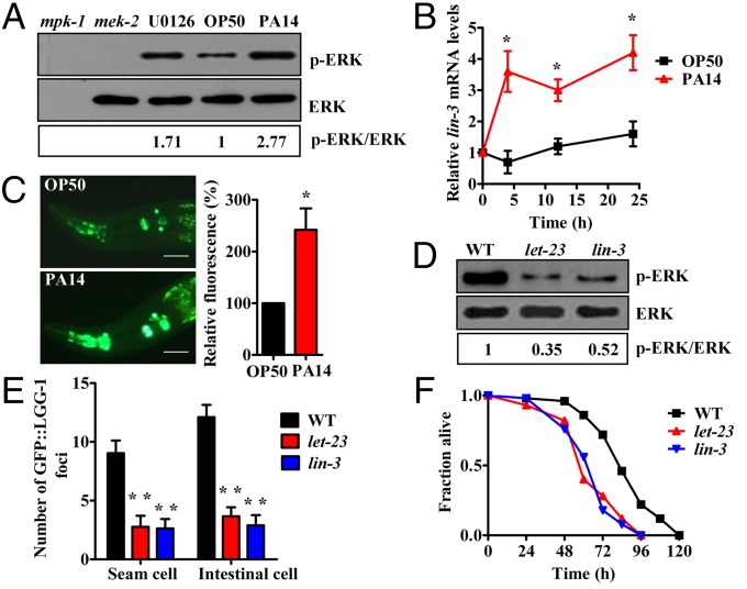
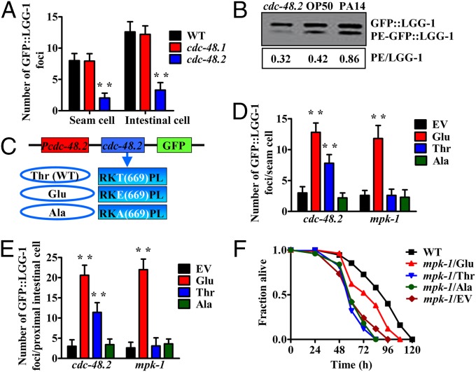
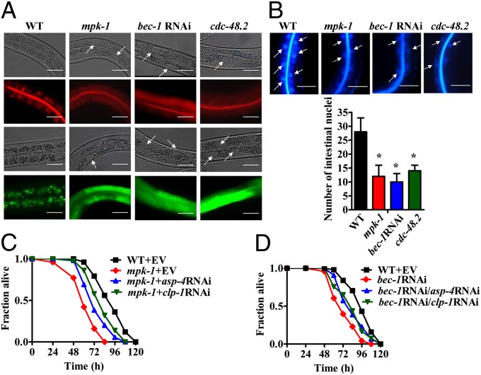
Autophagy, a conserved pathway that delivers intracellular materials into lysosomes for degradation, is involved in development, aging, and a variety of diseases. Accumulating evidence demonstrates that autophagy plays a protective role against infectious diseases by diminishing intracellular pathogens, including bacteria, viruses, and parasites. However, the mechanism by which autophagy regulates innate immunity remains largely unknown. Here, we show that autophagy is involved in host defense against a pathogenic bacterium Pseudomonas aeruginosa in the metazoan Caenorhabditis elegans. P. aeruginosa infection induces autophagy via a conserved extracellular signal-regulated kinase (ERK). Intriguingly, impairment of autophagy does not influence the intestinal accumulation of P. aeruginosa, but instead induces intestinal necrosis. Inhibition of necrosis results in the survival of autophagy-deficient worms after P. aeruginosa infection. These findings reveal a previously unidentified role for autophagy in protection against necrosis triggered by pathogenic bacteria in C. elegans and implicate that such a function of autophagy may be conserved through the inflammatory response in diverse organisms.
Conflict of interest statement
The authors declare no conflict of interest.
Figures
Similar articles
-
Translation initiation or elongation inhibition triggers contrasting effects on Caenorhabditis elegans survival during pathogen infection.mBio. 2024 Nov 13;15(11):e0248524. doi: 10.1128/mbio.02485-24. Epub 2024 Sep 30. mBio. 2024. PMID: 39347574 Free PMC article.
-
Pseudomonas aeruginosa suppresses host immunity by activating the DAF-2 insulin-like signaling pathway in Caenorhabditis elegans.PLoS Pathog. 2008 Oct;4(10):e1000175. doi: 10.1371/journal.ppat.1000175. Epub 2008 Oct 17. PLoS Pathog. 2008. PMID: 18927620 Free PMC article.
-
A conserved p38 MAP kinase pathway in Caenorhabditis elegans innate immunity.Science. 2002 Jul 26;297(5581):623-6. doi: 10.1126/science.1073759. Science. 2002. PMID: 12142542
-
Local and long-range activation of innate immunity by infection and damage in C. elegans.Curr Opin Immunol. 2016 Feb;38:1-7. doi: 10.1016/j.coi.2015.09.005. Epub 2015 Oct 28. Curr Opin Immunol. 2016. PMID: 26517153 Review.
-
Modeling Host-Pathogen Interactions in C. elegans: Lessons Learned from Pseudomonas aeruginosa Infection.Int J Mol Sci. 2024 Jun 27;25(13):7034. doi: 10.3390/ijms25137034. Int J Mol Sci. 2024. PMID: 39000143 Free PMC article. Review.
Cited by
-
Epithelial Atg5 Deficiency Intensifies Caspase-11 Activation, Fueling Extracellular mtDNA Release to Activate cGAS-STING-NLRP3 Axis in Macrophages During Pseudomonas Infection.MedComm (2020). 2025 Jun 15;6(7):e70239. doi: 10.1002/mco2.70239. eCollection 2025 Jul. MedComm (2020). 2025. PMID: 40529614 Free PMC article.
-
TOR functions as a molecular switch connecting an iron cue with host innate defense against bacterial infection.PLoS Genet. 2021 Mar 3;17(3):e1009383. doi: 10.1371/journal.pgen.1009383. eCollection 2021 Mar. PLoS Genet. 2021. PMID: 33657091 Free PMC article.
-
RNA-Sequencing of Heterorhabditis nematodes to identify factors involved in symbiosis with Photorhabdus bacteria.BMC Genomics. 2022 Nov 7;23(1):741. doi: 10.1186/s12864-022-08952-4. BMC Genomics. 2022. PMID: 36344922 Free PMC article.
-
Caenorhabditis elegans microRNAs of the let-7 family act in innate immune response circuits and confer robust developmental timing against pathogen stress.Proc Natl Acad Sci U S A. 2015 May 5;112(18):E2366-75. doi: 10.1073/pnas.1422858112. Epub 2015 Apr 20. Proc Natl Acad Sci U S A. 2015. PMID: 25897023 Free PMC article.
-
A unifying hypothesis on the central role of reactive oxygen species in bacterial pathogenesis and host defense in C. elegans.Curr Opin Immunol. 2021 Feb;68:9-20. doi: 10.1016/j.coi.2020.08.002. Epub 2020 Sep 6. Curr Opin Immunol. 2021. PMID: 32898751 Free PMC article. Review.
References
-
- Rubinsztein DC, Mariño G, Kroemer G. Autophagy and aging. Cell. 2011;146(5):682–695. - PubMed
-
- Nakagawa I, et al. Autophagy defends cells against invading group A Streptococcus. Science. 2004;306(5698):1037–1040. - PubMed
-
- Gutierrez MG, et al. Autophagy is a defense mechanism inhibiting BCG and Mycobacterium tuberculosis survival in infected macrophages. Cell. 2004;119(6):753–766. - PubMed
Publication types
MeSH terms
LinkOut - more resources
Full Text Sources
Other Literature Sources
Research Materials
Miscellaneous
