. 2014 Sep 23;111(38):13817-21.
doi: 10.1073/pnas.1408124111. Epub 2014 Aug 11.

A new tubulin-binding site and pharmacophore for microtubule-destabilizing anticancer drugs

Affiliations

A new tubulin-binding site and pharmacophore for microtubule-destabilizing anticancer drugs

Andrea E Prota et al. Proc Natl Acad Sci U S A. .

Abstract

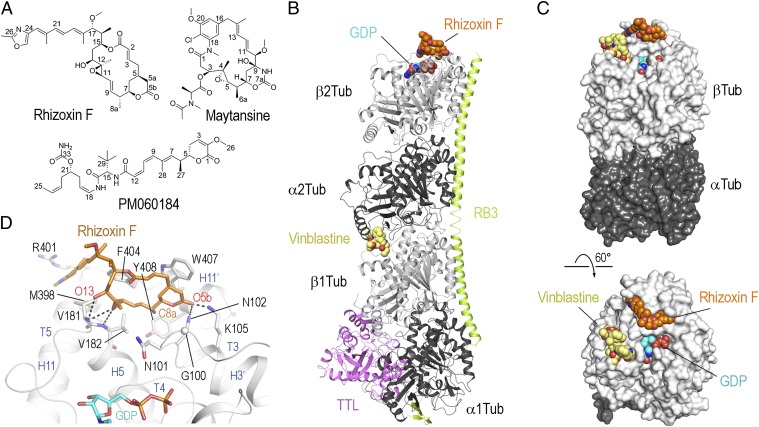
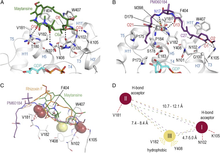
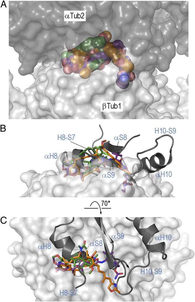
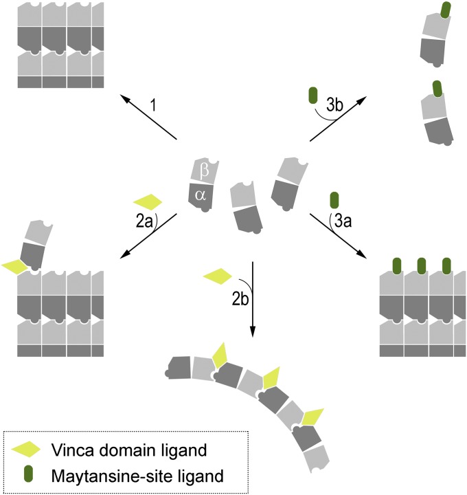
The recent success of antibody-drug conjugates (ADCs) in the treatment of cancer has led to a revived interest in microtubule-destabilizing agents. Here, we determined the high-resolution crystal structure of the complex between tubulin and maytansine, which is part of an ADC that is approved by the US Food and Drug Administration (FDA) for the treatment of advanced breast cancer. We found that the drug binds to a site on β-tubulin that is distinct from the vinca domain and that blocks the formation of longitudinal tubulin interactions in microtubules. We also solved crystal structures of tubulin in complex with both a variant of rhizoxin and the phase 1 drug PM060184. Consistent with biochemical and mutagenesis data, we found that the two compounds bound to the same site as maytansine and that the structures revealed a common pharmacophore for the three ligands. Our results delineate a distinct molecular mechanism of action for the inhibition of microtubule assembly by clinically relevant agents. They further provide a structural basis for the rational design of potent microtubule-destabilizing agents, thus opening opportunities for the development of next-generation ADCs for the treatment of cancer.

Keywords: X-ray crystallography; drug mechanism; microtubule-targeting agents.


Conflict of interest statement

The authors declare no conflict of interest.

Figures

Fig. 1.
Fig. 1.
Structure of the tubulin–rhizoxin F complex. (A) Chemical structures of rhizoxin F, maytansine, and PM060184. (B) Overall view of the T2R-TTL–rhizoxin F complex. Tubulin (gray), RB3 (light green), and TTL (violet) are shown in ribbon representation; the MDA rhizoxin F (orange) and GDP (cyan) are depicted in spheres representation. As a reference, the vinblastine structure (yellow, PDB ID no. 1Z2B) is superimposed onto the T2R complex. (C) Overall view of the tubulin–rhizoxin F interaction in two different orientations. The tubulin dimer with bound ligand (α-tubulin-2 and β-tubulin-2 of the T2R-TTL–rhizoxin F complex) is shown in surface representation. The vinblastine structure is superimposed onto the β-tubulin chain to highlight the distinct binding site of rhizoxin F. All ligands are in sphere representation and are colored in orange (rhizoxin F), cyan (GDP), and yellow (vinblastine). (D) Close-up view of the interaction observed between rhizoxin F (orange sticks) and β-tubulin (gray ribbon). Interacting residues of β-tubulin are shown in stick representation and are labeled.
Fig. 2.
Fig. 2.
Structures of the tubulin–maytansine and tubulin–PM060184 complexes and pharmacophore model. (A) Close-up view of the tubulin–maytansine complex. Maytansine is in green stick representation. β-tubulin is displayed as gray ribbon. Key residues forming the interaction with the ligand are in stick representation and are labeled. Hydrogen bonds are highlighted as dashed black lines. (B) Detailed view of the tubulin–PM060184 complex. The ligand is displayed as violet-purple sticks. (C) Superposition of the binding sites of rhizoxin F (orange), maytansine (green), and PM060184 (violet-purple) highlighting the three common interaction points I, II, and III with β-tubulin. Hydrogen bond acceptors are highlighted as red spheres; the methyl groups forming the hydrophobic interaction are highlighted as yellow spheres. (D) Schematic drawing of the common pharmacophore for ligand binding to the maytansine site, using the same color code as in C.
Fig. 3.
Fig. 3.
Binding of maytansine-site ligands in the context of a microtubule. (A) View of the tubulin–maytansine-site ligand interaction in the context of the microtubule (PDB ID no. 2XRP). The binding sites of the complexes of rhizoxin F, maytansine, and PM060184 are superimposed on the corresponding site on β-tubulin of the microtubule model. The α- and β-tubulin chains are displayed as dark and light gray surfaces, respectively. The ligands are in sphere representation, using the same color code as in Figs. 1C and 2 A and B. (B) Side view of the longitudinal tubulin–tubulin contact with superimposed maytansine-site ligands. For clarity reasons, only the secondary structure elements of α-tubulin forming the longitudinal contact are shown and labeled in light blue. β-Tubulin is in gray surface; the maytansine-site ligands are in stick representation. (C) Top view of the longitudinal tubulin–tubulin contact highlighting the prominent steric clash between the maytansine-site ligands bound to β-tubulin and the helix H8 of α-tubulin from a neighboring dimer. The same settings as in B are used.
Fig. 4.
Fig. 4.
Molecular mechanism of action of vinca domain- and maytansine-site ligands on tubulin and microtubules. (1) In the absence of ligands, curved αβ-tubulin heterodimers assemble into microtubules and undergo a curved-to-straight conformational transition. Formation of longitudinal contacts include the interaction between a pocket shaped by loops S3-H3, S5-H5, and H11-H11′ of the β-tubulin subunit of one dimer (cavity) and helix H8 of α-tubulin from a neighboring dimer in the microtubule lattice (knob). (2) Vinblastine binds to the vinca domain, a composite binding site that is formed by structural elements from both α- and β-tubulin monomers of two different, longitudinally aligned αβ-tubulin heterodimers. The MDA destabilizes microtubules by introducing a wedge at the interface between two tubulin dimers at the tips of microtubules, thus inhibiting the curved-to-straight conformational transition necessary to build up the microtubule lattice (2a), or by stabilizing curved, ring-like oligomers that are not compatible with the straight tubulin structure found in microtubules (2b). (3) Maytansine-site ligands bind to the site on β-tubulin that is involved in the formation of longitudinal contacts in microtubules. These types of MDAs destabilize microtubules either by binding to the plus ends of growing microtubules at substoichiometric ligand concentrations, thus inhibiting the addition of further tubulin subunits (3a), or by forming assembly incompetent tubulin–drug complexes with unassembled tubulin subunits at high ligand concentrations (3b). Note that in vitro at high Mg2+ concentrations, tubulin–PM060184 complexes can assemble into small ring-like oligomers (14).

Comment in

  • A previously undescribed tubulin binder.
    Field JJ, Waight AB, Senter PD. Field JJ, et al. Proc Natl Acad Sci U S A. 2014 Sep 23;111(38):13684-5. doi: 10.1073/pnas.1414572111. Epub 2014 Sep 3. Proc Natl Acad Sci U S A. 2014. PMID: 25187564 Free PMC article. No abstract available.

References

    1. Dumontet C, Jordan MA. Microtubule-binding agents: A dynamic field of cancer therapeutics. Nat Rev Drug Discov. 2010;9(10):790–803. - PMC - PubMed
    1. Jordan MA, Wilson L. Microtubules as a target for anticancer drugs. Nat Rev Cancer. 2004;4(4):253–265. - PubMed
    1. Canta A, Chiorazzi A, Cavaletti G. Tubulin: A target for antineoplastic drugs into the cancer cells but also in the peripheral nervous system. Curr Med Chem. 2009;16(11):1315–1324. - PubMed
    1. Zolot RS, Basu S, Million RP. Antibody-drug conjugates. Nat Rev Drug Discov. 2013;12(4):259–260. - PubMed
    1. Teicher BA, Doroshow JH. The promise of antibody-drug conjugates. N Engl J Med. 2012;367(19):1847–1848. - PubMed

Publication types

MeSH terms