. 2014:2014:701094.
doi: 10.1155/2014/701094. Epub 2014 Jul 8.

Overexpression and selectively regulatory roles of IL-23/IL-17 axis in the lesions of oral lichen planus

Affiliations

Overexpression and selectively regulatory roles of IL-23/IL-17 axis in the lesions of oral lichen planus

Rui Lu et al. Mediators Inflamm. 2014.

Abstract

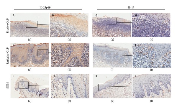
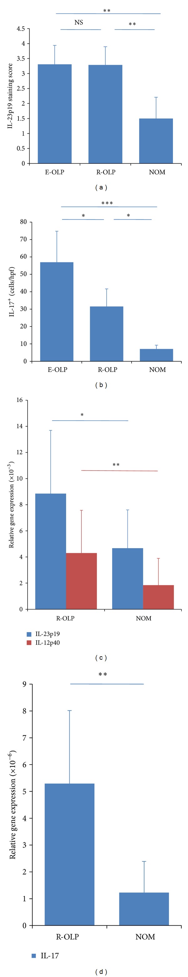
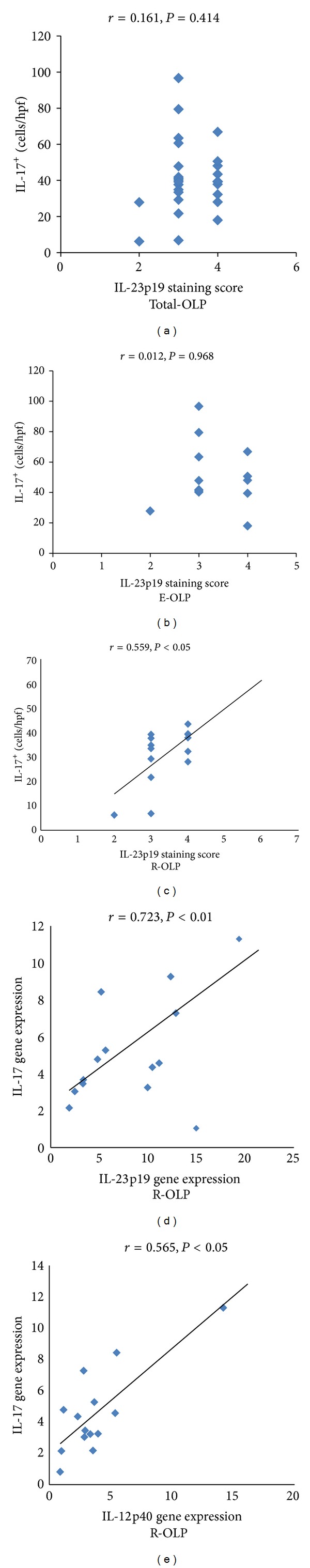
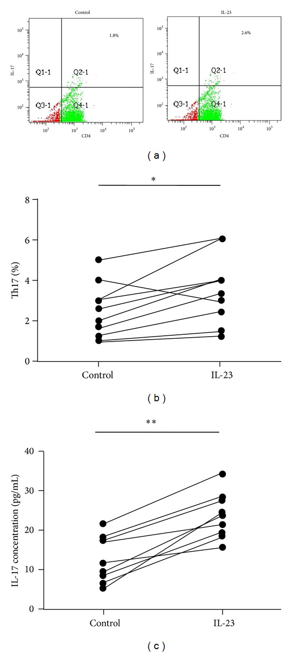
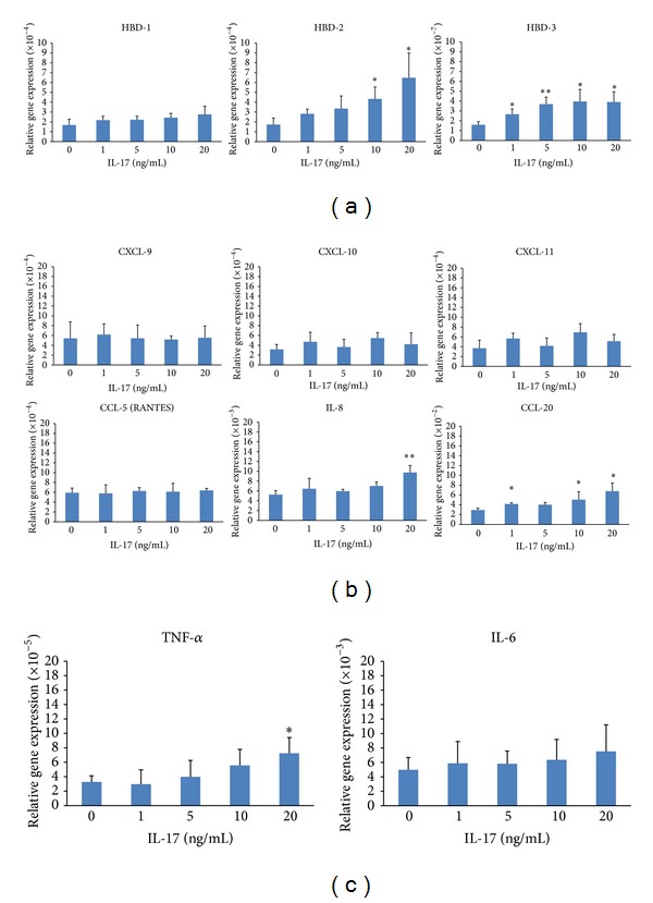
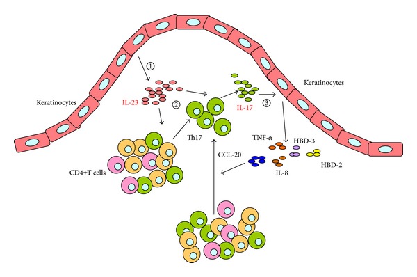
Interleukin- (IL-) 23/IL-17 axis is a newly discovered proinflammatory signaling pathway and has been implicated in the pathogenesis of many chronic inflammatory and immune disorders. Here we investigated whether the IL-23/IL-17 axis was present and functional in the lesions of oral lichen planus (OLP), a chronic inflammatory disease affecting the oral mucosa. Using immunohistochemistry and quantitative PCR, we found that the subunits of IL-23 and IL-17 were overexpressed in OLP lesions than in normal oral mucosa tissues. In addition, the expressions of IL-23 and IL-17 are positively correlated in reticular OLP tissues. Results from in vitro studies revealed that exogenous IL-23 could increase the percentage of Th17 cells and IL-17 production in the CD4+T cells from reticular OLP patients. Furthermore, we also found that exogenous IL-17 could significantly enhance the mRNA expressions of β-defensin-2, -3, CCL-20, IL-8, and TNF-α, but not β-defensin-1, CXCL-9, -10, -11, CCL-5, and IL-6 in human oral keratinocytes. Taken together, our results revealed an overexpression pattern and selectively regulatory roles of IL-23/IL-17 axis in the OLP lesions, suggesting that it may be a pivotal regulatory pathway in the complex immune network of OLP lesions.


Figures

Figure 1
Figure 1
Immunohistochemical stainings for IL-23p19 (a–f) and IL-17 (g–l) in erosive (a, b, g, and h) and reticular (c, d, i, and j) OLP lesions and normal oral mucosa tissues (e, f, k, and l). Immunohistochemical staining for IL-23p19 showed diffuse and strong patterns in epithelium and the extracellular matrix of the lamina propria of both erosive ((a) ×100; (b) ×400) and reticular ((c) ×100; (d) ×400) OLP lesions, but weak or absent pattern in normal oral mucosa tissues ((e) ×100; (f) ×400). Abundant IL-17 positive staining was observed on the cytoplasm of the infiltrated lymphocytes in the lesions of both erosive ((g) ×100; (h) ×400) and reticular ((i) ×100; (j) ×400) OLP, but only a few sporadic IL-17+ cells were seen in normal oral mucosa ((k) ×100; (l) ×400).
Figure 2
Figure 2
Expressions of IL-23 and IL-17 in OLP lesions. (a) The average staining scores of IL-23p19 in erosive OLP lesions (n = 13), reticular OLP lesions (n = 14), and normal oral mucosa tissues (n = 10). (b) The average number of IL-17+ cells per hpf in erosive OLP lesions (n = 13), reticular OLP lesions (n = 14), and normal oral mucosa tissues (n = 10). ((c) and (d)) The mRNA expressions of IL-23p19, IL-12p40, and IL-17 in reticular OLP lesions (n = 14) and normal oral mucosa tissues (n = 10). All data were shown as mean ± SEM. **P < 0.01; **P < 0.05; NS: nonsignificantly.
Figure 3
Figure 3
Correlation between the expressions of IL-23 and IL-17 in OLP tissue specimens. (a–c) Correlations between the staining scores of IL-23p19 and numbers of IL-17+ cells per hpf in total OLP tissue specimens (n = 27, r = 0.161, P > 0.05), erosive OLP tissue specimens (n = 13, r = 0.012, P > 0.05) and reticular OLP tissue specimens (n = 14, r = 0.559, P < 0.05). (d-e) Correlations between the mRNA expressions of IL-23p19 ((d) r = 0.723, P < 0.01) or IL-12p40 ((e) r = 0.565, P < 0.05) and IL-17 in reticular OLP tissue specimens (n = 14).
Figure 4
Figure 4
The effect of recombinant (r) IL-23 on the percentage of Th17 cells and IL-17 production in CD4+T cells from OLP patients. (a) Representative scatter plots of CD4+IL-17+ staining in peripheral blood CD4+T cells from OLP patients (n = 10), with or without the stimulation of rIL-23 (20 ng/mL) for 36 h. ((b) and (c)) Paired comparisons of percentages of Th17 cells (b) and the IL-17 content in the culture supernatant (c) in peripheral blood CD4+T cells from OLP patients (n = 10), with or without the stimulation of rIL-23 (20 ng/mL) for 36 h.
Figure 5
Figure 5
The effect of recombinant (r) IL-17 to the mRNA expressions of inflammatory mediators, including β-defensins (a), chemokines (b), and proinflammatory cytokines (c), on the HOK cells. Results were shown as mean ± SEM. **P < 0.01; **P < 0.05.
Figure 6
Figure 6
Schematic model of IL-23/IL-17 axis involved in the pathogenesis of OLP. The whole process is divided into three steps: (1) keratinocytes in OLP lesion produce a large amount of IL-23 via an unknown mechanism; (2) keratinocyte-derived IL-23 may contribute to the accumulation of Th17 cells and the overproduction of IL-17 in the local lesion of OLP; (3) IL-17 reversely induces the keratinocytes to selectively produce various inflammatory mediators, which compose the complex immune network in the inflammatory environment of OLP lesions.

References

    1. Lodi G, Scully C, Carrozzo M, Griffiths M, Sugerman PB, Thongprasom K. Current controversies in oral lichen planus: report of an international consensus meeting. Part 1. Viral infections and etiopathogenesis. Oral Surgery, Oral Medicine, Oral Pathology, Oral Radiology and Endodontology. 2005;100(1):40–51. - PubMed
    1. Sugerman PB, Savage NW, Walsh LJ, et al. The pathogenesis of oral lichen planus. Critical Reviews in Oral Biology and Medicine. 2002;13(4):350–365. - PubMed
    1. Eisen D, Carrozzo M, Sebastian J-VB, Thongprasom K. Number V. Oral lichen planus: clinical features and management. Oral Diseases. 2005;11(6):338–349. - PubMed
    1. Roopashree MR, Gondhalekar RV, Shashikanth MC, George J, Thippeswamy SH, Shukla A. Pathogenesis of oral lichen planus—a review. Journal of Oral Pathology and Medicine. 2010;39(10):729–734. - PubMed
    1. Lavanya N, Jayanthi P, Rao U, Ranganathan K. Oral lichen planus: an update on pathogenesis and treatment. Journal of Oral and Maxillofacial Pathology. 2011;15(2):127–132. - PMC - PubMed

Publication types

LinkOut - more resources