

Case Reports
doi: 10.4103/0971-3026.137053.
Morel-Lavallée lesion: A closed degloving injury that requires real attention
Affiliations
- PMID: 25114393
- PMCID: PMC4126145
- DOI: 10.4103/0971-3026.137053
Item in Clipboard
Case Reports
Morel-Lavallée lesion: A closed degloving injury that requires real attention
Indian J Radiol Imaging.
2014 Jul.
Abstract
Morel-Lavallée lesions are post-traumatic, closed degloving injuries occurring deep to subcutaneous plane due to disruption of capillaries resulting in an effusion containing hemolymph and necrotic fat. Magnetic resonance imaging (MRI) is the modality of choice in the evaluation of Morel-Lavallée lesion. Early diagnosis and management is essential as any delay in diagnosis or missed lesion will lead to the effusion becoming infected or leading to extensive skin necrosis.
Keywords: Closed degloving injury; Morel-Lavallée lesions; magnetic resonance imaging.
Conflict of interest statement
Figures
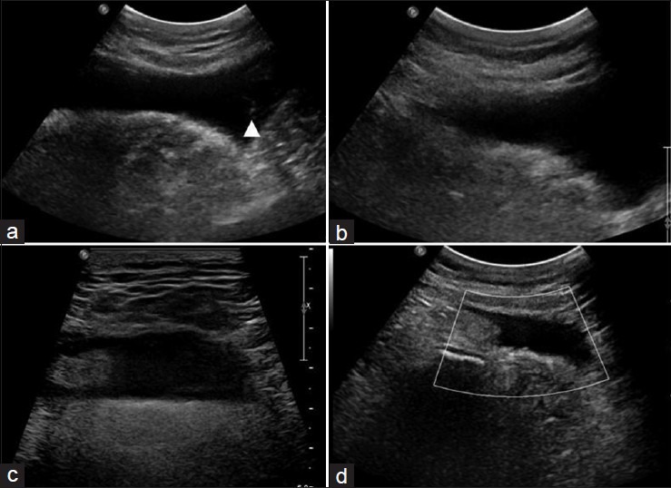
USG scan of fluctuant swelling in left anterolateral thigh showing, (a) large complex septated (arrow head) fluid collection within deep subcutaneous tissues; (b and c) fluid collection with internal echoes; (d) no evidence of vascularity
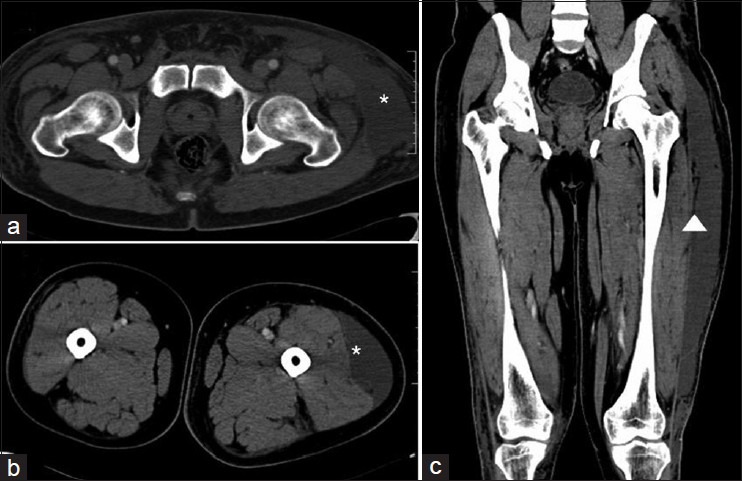
CT axial images (a and b) hypodense collection (asterisk) within deep subcutaneous tissues; Coronal image (c) streaks of fat density (arrow head) within subcutaneous collection
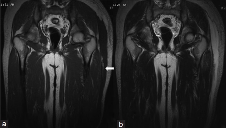
MR (a) Coronal T1W image showing, subcutaneous hypointense-fluid collection with fat density (arrow); (b) Coronal T2W image, showing hyperintense subcutaneous fluid collection
References
-
- Letournel E, Judet R. 2nd ed. Berlin, Germany: Springer-Verlag; 1993. Fractures of the acetabulum.
-
- Hak DJ, Olson SA, Matta JM. Diagnosis and management of closed internal degloving injuries associated with pelvic and acetabular fractures: The Morel-Lavallée lesion. J Trauma. 1997;42:1046–51. - PubMed
-
- Kottmeier SA, Wilson SC, Born CT, Hanks GA, Iannacone WM, Delong WG. Surgical management of soft tissue lesions associated with pelvic ring injury. Clin Orthop Relat Res. 1996;329:46–53. - PubMed
-
- Parra JA, Fernandez MA, Encinas B, Rico M. Morel-Lavallée effusions in the thigh. Skeletal Radiol. 1997;26:239–41. - PubMed
-
- Rubin JI, Gomori JM, Grossman RI, Gefter WB, Kressel HY. High-field MR imaging of extracranial hematomas. AJR Am J Roentgenol. 1987;148:813–7. - PubMed
Publication types
LinkOut - more resources
Full Text Sources
Other Literature Sources