

Case Reports
doi: 10.1007/s12288-012-0206-3.
Epub 2012 Nov 6.
Pseudo chediak-higashi granules in acute lymphoblastic leukemia: a rare entity
Affiliations
- PMID: 25114409
- PMCID: PMC4115082
- DOI: 10.1007/s12288-012-0206-3
Item in Clipboard
Case Reports
Pseudo chediak-higashi granules in acute lymphoblastic leukemia: a rare entity
Indian J Hematol Blood Transfus.
2014 Sep.
Abstract
Pseudo-Chediak-Highashi granules are giant cytoplasmic inclusions commonly encountered in myeloblasts or other myeloid precursors in acute myeloid leukemia and myelodysplastic syndromes. They derive their name from the inherited Chediak-Higashi syndrome that presents with oculocutaneous albinism, chronic infections and platelet dense granule deficiency. We report possibly the third case in world literature where these granules were seen in the blast cells of acute lymphoblastic leukemia in a 15-year-old male.
Keywords: Acute lymphoblastic leukemia; Bone marrow; Flow cytometry; Morphology; Pseudo Chediak-Highashi granules.
Figures
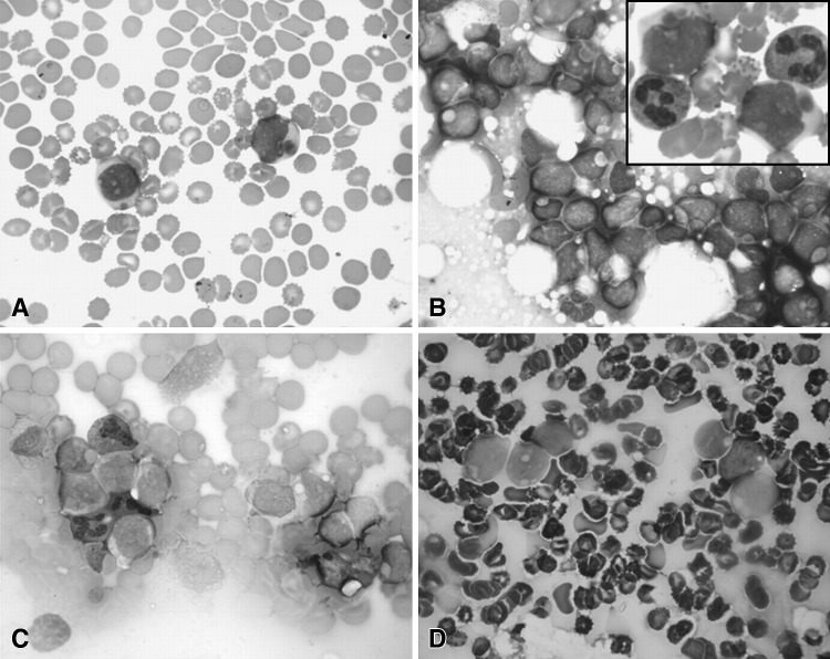
Peripheral smear shows blasts with pseudo Chediak-Higashi granules (MGG-Giemsa stain, ×1000, a). Bone marrow aspirate shows many blasts with similar large granules. (MGG-Giemsa stain, ×1000 & Inset, b). PAS stain shows faint positivity for these blasts (PAS ×1000, c) whiles these cells are negative for MPO (MPO ×1000, d)
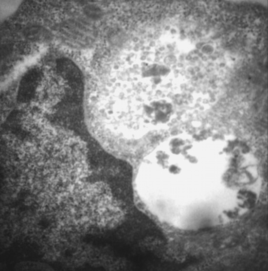
Ultrastructural examination shows presence of perinuclear membrane bound structure. These structures contain multiple uniform-sized vesicles lined by a unit membrane (EM ×10800)
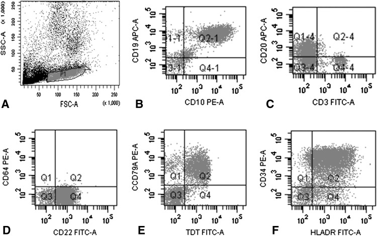
Scatter plots depict multicolor immunophenotyping on bone marrow aspirate by flow cytometry. The events are seen on side scatter (SSC) versus forward scatter (FSC) plot. The cells in blastic region are gated as P1 and the same cells are analysed in subsequent plots (a). These cells show dual bright expression of CD19 and CD10 (b) along with dim expression of CD 20 (c). The same cell populations show expression CD22 (d). These cell show expression of CD79a, TdT, CD34 and HLA-DR (e, f)
References
-
- Tsai IM, Tsai CC, Ladd DJ. Pseudo-Chediak-Higashi anomaly in chronic myelogenous leukemia with myelofibrosis. Am J Clin Pathol. 1977;67(6):608–609. - PubMed
Publication types
LinkOut - more resources
Full Text Sources
Research Materials